Fundamentals of the Nervous System: Nervous Tissue Structure and Cell Functions
Lesson Overview
- What Is the Nervous System and Why Is It Essential for Human Functioning?
- How Is the Nervous System Functionally Organized?
- What Are Neurons and How Do They Transmit Signals?
- How Are Neurons Classified Based on Structure and Function?
- What Is the Role of Neuroglia in the Nervous System?
- What Is Myelination and How Does It Enhance Signal Transmission?
- What Is the Resting Membrane Potential and How Is It Maintained?
- What Are Graded Potentials and Action Potentials?
- How Do Gated Channels Control Ion Movement?
- What Happens at the Synapse During Signal Transmission?
- What Are the Differences Between the Somatic and Autonomic Nervous Systems?
- How Do Afferent and Efferent Fibers Function?
When students struggle to understand how the brain controls everything from reflexes to emotions, frustration quickly builds up. This lesson on the fundamentals of the nervous system and nervous tissue breaks down that complexity. It offers a clear, structured way to grasp how neural signals work, making the subject much easier to learn.
What Is the Nervous System and Why Is It Essential for Human Functioning?
The nervous system is the body's master communication and control center. It integrates and coordinates all body activities by transmitting electrical and chemical signals. Every thought, emotion, sensation, and action originates within the nervous system. Understanding its organization and components is crucial for grasping how the body functions.
- The nervous system is responsible for maintaining homeostasis, initiating movements, and interpreting sensory information.
- It detects changes (stimuli) in the internal and external environment and responds accordingly.
- It enables higher cognitive functions like learning, memory, and language.
The nervous system consists of two main structural divisions:
| Division | Components | Primary Function |
| Central Nervous System (CNS) | Brain and Spinal Cord | Integration and processing of information |
| Peripheral Nervous System (PNS) | Cranial and spinal nerves | Communication between CNS and body |
The CNS acts as the command center, while the PNS serves as the communication network connecting the CNS to the rest of the body.
(161).jpg)
How Is the Nervous System Functionally Organized?
The nervous system performs three interconnected functions: sensory input, integration, and motor output.
- Sensory input involves detecting stimuli through receptors located in skin, muscles, joints, and organs.
- Integration refers to processing and interpreting sensory input to determine the appropriate response.
- Motor output activates effector organs like muscles and glands to produce a response.
Functionally, the PNS is divided into two major divisions:
| Functional Division | Direction of Signal | Description |
| Sensory (Afferent) Division | To CNS | Carries information from sensory receptors to the CNS |
| Motor (Efferent) Division | From CNS | Transmits commands from CNS to muscles and glands |
The motor division includes:
- The somatic nervous system, which controls voluntary movements.
- The autonomic nervous system, which regulates involuntary activities like heartbeat and digestion.
What Are Neurons and How Do They Transmit Signals?
Neurons are specialized, excitable cells capable of generating and transmitting electrical impulses called action potentials. They form the basic structural and functional units of the nervous system.
Each neuron has the following structures:
- Cell body (soma): Contains the nucleus and metabolic machinery.
- Dendrites: Short, branched extensions that receive incoming signals.
- Axon: A long extension that transmits signals away from the soma.
- Axon terminals: End structures that release neurotransmitters.
Neurons rely on electrical gradients and ion channels to propagate impulses. They communicate through both electrical and chemical synapses.
| Neuron Part | Function |
| Soma | Metabolism and integration of incoming signals |
| Dendrites | Receive electrical stimuli from other neurons |
| Axon | Conducts action potentials to target cells |
| Axon terminals | Release neurotransmitters into the synaptic cleft |
How Are Neurons Classified Based on Structure and Function?
Structural classification is based on the number of processes extending from the soma:
- Multipolar neurons have one axon and multiple dendrites; most common in the CNS.
- Bipolar neurons have one dendrite and one axon; found in the retina and olfactory mucosa.
- Unipolar neurons have a single short process that splits into two branches; found in sensory ganglia of the PNS.
| Type | Processes | Example Locations |
| Multipolar | One axon, many dendrites | Brain, spinal cord (motor neurons) |
| Bipolar | One axon, one dendrite | Retina, inner ear, olfactory epithelium |
| Unipolar | Single short process | Dorsal root ganglia |
Functionally, neurons are classified into:
- Sensory (afferent) neurons: Transmit sensory input to the CNS.
- Motor (efferent) neurons: Carry signals from the CNS to effectors.
- Interneurons: Integrate sensory input and coordinate motor output within the CNS.
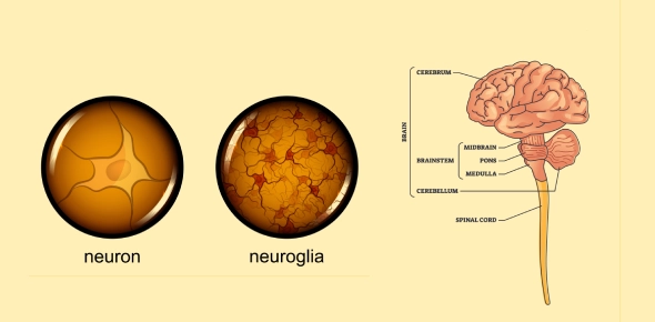
What Is the Role of Neuroglia in the Nervous System?
Neuroglia are non-excitable support cells that vastly outnumber neurons. They provide structural support, insulation, nutrition, and immune protection.
| Glial Cell | Location | Functions |
| Astrocytes | CNS | Regulate ion balance, support neurons, maintain blood-brain barrier |
| Microglia | CNS | Act as phagocytes, remove debris, defend against pathogens |
| Ependymal cells | CNS | Produce and circulate cerebrospinal fluid (CSF) |
| Oligodendrocytes | CNS | Form myelin sheath in CNS axons |
| Schwann cells | PNS | Form myelin sheath in PNS axons |
| Satellite cells | PNS | Surround neuron cell bodies in ganglia, regulate environment |
These cells ensure optimal function of the nervous system and play a role in regeneration and response to injury.
What Is Myelination and How Does It Enhance Signal Transmission?
Myelin is a lipid-rich insulating material that wraps around axons, formed by oligodendrocytes in the CNS and Schwann cells in the PNS. It increases conduction velocity by allowing saltatory conduction.
- Saltatory conduction involves action potentials leaping between nodes of Ranvier.
- Myelination reduces energy expenditure and enhances speed.
- Damage to myelin leads to diseases such as multiple sclerosis.
| Region | Structure | Composition |
| White Matter | Myelinated axons | Rich in lipids (myelin) |
| Gray Matter | Neuron cell bodies, dendrites | Low myelin content |
What Is the Resting Membrane Potential and How Is It Maintained?
The resting membrane potential is the voltage difference between the inside and outside of a resting neuron, typically about -70 mV.
- It arises due to the distribution of Na+, K+, and Cl- ions across the membrane.
- Potassium ions are more concentrated inside the cell, while sodium ions are more concentrated outside.
- The Na+-K+ pump expels 3 Na+ and brings in 2 K+ per cycle, maintaining this difference.
This potential is essential for the generation of action potentials.
What Are Graded Potentials and Action Potentials?
Graded potentials are local, variable-strength signals that decay over distance. They result from the opening of chemically gated channels in dendrites or soma.
If a graded potential reaches threshold (-55 mV), it triggers an action potential at the axon hillock.
Action potentials are uniform, all-or-none electrical signals that travel along axons.
| Feature | Graded Potential | Action Potential |
| Strength | Varies with stimulus | Fixed amplitude |
| Duration | Short | Longer |
| Decay | Yes | No |
| Channels Involved | Ligand-gated | Voltage-gated |
How Do Gated Channels Control Ion Movement?
Gated ion channels regulate ion flow across membranes and are essential for excitability.
- Chemically gated channels open in response to neurotransmitters.
- Voltage-gated channels open when membrane potential changes.
- Mechanically gated channels open due to physical deformation.
Opening these channels allows specific ions to flow, generating electrical currents and changing membrane potential.
What Happens at the Synapse During Signal Transmission?
A synapse is a junction where a neuron communicates with another cell.
- The presynaptic neuron releases neurotransmitters from vesicles.
- These chemicals diffuse across the synaptic cleft.
- The postsynaptic neuron binds neurotransmitters via receptors, initiating a graded potential.
| Synaptic Type | Description |
| Axodendritic | Axon to dendrite |
| Axosomatic | Axon to soma |
| Axoaxonic | Axon to axon |
Neurotransmitter release is calcium-dependent and ensures one-way transmission of signals.
What Are the Differences Between the Somatic and Autonomic Nervous Systems?
The motor division of the nervous system splits into:
| Feature | Somatic Nervous System | Autonomic Nervous System |
| Control | Voluntary | Involuntary |
| Effectors | Skeletal muscles | Smooth, cardiac muscles, glands |
| Neurons in Pathway | One | Two (pre and post-ganglionic) |
The autonomic nervous system further divides into:
- Sympathetic division: Mobilizes energy, increases heart rate.
- Parasympathetic division: Conserves energy, lowers heart rate.
How Do Afferent and Efferent Fibers Function?
Afferent (sensory) neurons transmit information to the CNS.
- Somatic afferents come from skin, muscles, and joints.
- Visceral afferents come from internal organs.
Efferent (motor) neurons convey signals from the CNS to effector tissues:
- Somatic motor neurons stimulate skeletal muscles.
- Autonomic motor neurons influence glands, cardiac, and smooth muscles.
Rate this lesson:
 Back to top