PEDS Cardio

24 Questions | Attempts: 250
Share

SettingsSettingsSettings
PEDS Cardio - Quiz

Questions from small groups to help prep for course 3 exam


Questions and Answers
  • 1. 

    A 4-week old girl presents with poor feeding and tachypnea.  The infant was born at term after a normal pregnancy.  At 2 weeks of ago, a cardiac murmur was noted, but the baby was otherwise well.  However, over the last week she has become more tachypneic and is feeding poorly.  Her family is concerned that she takes up to 50 minutes to feed and becomes sweaty and tachypneic during feedings.  There is no history of cough or fever.  What physiological change takes place at approximately 4 weeks of age that could be important to consider in this case?

    • A.

      Closing of ductus arteriosus

    • B.

      Closing of foramen ovale

    • C.

      Closing of ductus venosus

    • D.

      Decline in pulmonary vascular resistance

    Correct Answer
    D. Decline in pulmonary vascular resistance
    Explanation
    closing of foramen ovale occurs at, or shortly after, birth. Closing of ductus arteriosus and ductus venosus occur shortly after birth. The decline in pulmonary vascular resistance can take a few weeks.

    Rate this question:

  • 2. 

    A 4-week old girl presents with poor feeding and tachypnea.  The infant was born at term after a normal pregnancy.  At 2 weeks of ago, a cardiac murmur was noted, but the baby was otherwise well.  However, over the last week she has become more tachypneic and is feeding poorly.  Her family is concerned that she takes up to 50 minutes to feed and becomes sweaty and tachypneic during feedings.  There is no history of cough or fever.  On physical exam, weight 3.2kg (>5th %ile), length 53cm (~25th %ile), temp 36.5, HR 160, RR 64, BP (right arm) 80/40, O2 sat 92%.  She is pale and diaphoretic.  There is subcostal indrawing.  The cardiac apex is just lateral to the mid-clavicular line at the 5th left intercostal space, and the precordial activity is increased.  The heart sounds are normal, a harsh 3/6 pansystolic murmur is present.  The liver is 4cm below the right costal margin.  All of the following are causes of a pansystolic murmur EXCEPT

    • A.

      Ventral septal defect

    • B.

      Mitral valve regurgitation

    • C.

      Pulmonary stenosis

    • D.

      Tricuspid valve stenosis

    Correct Answer
    C. Pulmonary stenosis
  • 3. 

    A 4-week old girl presents with poor feeding and tachypnea.  The infant was born at term after a normal pregnancy.  At 2 weeks of ago, a cardiac murmur was noted, but the baby was otherwise well.  However, over the last week she has become more tachypneic and is feeding poorly.  Her family is concerned that she takes up to 50 minutes to feed and becomes sweaty and tachypneic during feedings.  There is no history of cough or fever.  On physical exam, weight 3.2kg (>5th %ile), length 53cm (~25th %ile), temp 36.5, HR 160, RR 64, BP (right arm) 80/40, O2 sat 92%.  She is pale and diaphoretic.  There is subcostal indrawing.  The cardiac apex is just lateral to the mid-clavicular line at the 5th left intercostal space, and the precordial activity is increased.  The heart sounds are normal, a harsh 3/6 pansystolic murmur is present.  The liver is 4cm below the right costal margin.Where should the pediatric cardiac apex be?

    • A.

      3rd intercostal space

    • B.

      4th intercostal space

    • C.

      5th intercostal space

    • D.

      6th intercostal space

    Correct Answer
    B. 4th intercostal space
  • 4. 

    A 4-week old girl presents with poor feeding and tachypnea.  The infant was born at term after a normal pregnancy.  At 2 weeks of ago, a cardiac murmur was noted, but the baby was otherwise well.  However, over the last week she has become more tachypneic and is feeding poorly.  Her family is concerned that she takes up to 50 minutes to feed and becomes sweaty and tachypneic during feedings.  There is no history of cough or fever.  On physical exam, weight 3.2kg (>5th %ile), length 53cm (~25th %ile), temp 36.5, HR 160, RR 64, BP (right arm) 80/40, O2 sat 92%.  She is pale and diaphoretic.  There is subcostal indrawing.  The cardiac apex is just lateral to the mid-clavicular line at the 5th left intercostal space, and the precordial activity is increased.  The heart sounds are normal, a harsh 3/6 pansystolic murmur is present.  The liver is 4cm below the right costal margin.  Are you concerned about hepatomegaly?

    • A.

      Yes

    • B.

      No

    • C.

      Splenectomy!

    Correct Answer
    A. Yes
    Explanation
    yes, 4cm below is ENORMOUS considering total torso size of a 4-week old baby!

    Rate this question:

  • 5. 

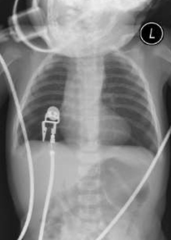
    A 4-week old girl presents with poor feeding and tachypnea.  The infant was born at term after a normal pregnancy.  At 2 weeks of ago, a cardiac murmur was noted, but the baby was otherwise well.  However, over the last week she has become more tachypneic and is feeding poorly.  Her family is concerned that she takes up to 50 minutes to feed and becomes sweaty and tachypneic during feedings.  There is no history of cough or fever.  On physical exam, weight 3.2kg (>5th %ile), length 53cm (~25th %ile), temp 36.5, HR 160, RR 64, BP (right arm) 80/40, O2 sat 92%.  She is pale and diaphoretic.  There is subcostal indrawing.  The cardiac apex is just lateral to the mid-clavicular line at the 5th left intercostal space, and the precordial activity is increased.  The heart sounds are normal, a harsh 3/6 pansystolic murmur is present.  The liver is 4cm below the right costal margin.     This CXR shows all of the following EXCEPT

    • A.

      Cardiomegaly

    • B.

      Increased pulmonary vascularity

    • C.

      Mild pulmonary edema

    • D.

      NG tube

    • E.

      Tracheal deviation

    Correct Answer
    E. Tracheal deviation
    Explanation
    the CXR shows mild cardiomegaly with increased pulmonary vascularity, and mild pulmonary edema. Bone and soft tissues are normal. There is a nasogastric tube in stomach.

    Rate this question:

  • 6. 

    Which of the following is seen in this ECG (check all that apply) 

    • A.

      Sinus tachycardia

    • B.

      Right ventricular hypertrophy

    • C.

      Left ventricular hypertrophy

    • D.

      Abnormal T waves

    • E.

      Normal axis

    Correct Answer(s)
    A. Sinus tachycardia
    B. Right ventricular hypertrophy
    C. Left ventricular hypertrophy
    E. Normal axis
    Explanation
    sinus tachycardia (HR 150-160 bpm) and biventricular hypertrophy (large voltages throughout the precordial leads). Upright T wave in v6 mandatory, inverted T waves in v1-v3 are appropriate. V1 will be the last one to flip upright, well into puberty. Normal baby axis between II and II (R heart dominant)

    Rate this question:

  • 7. 

    A 4-week old girl presents with poor feeding and tachypnea.  The infant was born at term after a normal pregnancy.  At 2 weeks of ago, a cardiac murmur was noted, but the baby was otherwise well.  However, over the last week she has become more tachypneic and is feeding poorly.  Her family is concerned that she takes up to 50 minutes to feed and becomes sweaty and tachypneic during feedings.  There is no history of cough or fever.  On physical exam, weight 3.2kg (>5th %ile), length 53cm (~25th %ile), temp 36.5, HR 160, RR 64, BP (right arm) 80/40, O2 sat 92%.  She is pale and diaphoretic.  There is subcostal indrawing.  The cardiac apex is just lateral to the mid-clavicular line at the 5th left intercostal space, and the precordial activity is increased.  The heart sounds are normal, a harsh 3/6 pansystolic murmur is present.  The liver is 4cm below the right costal margin.  Is this a cyanotic lesion?

    • A.

      Yes

    • B.

      No

    • C.

      Hold on, I'll check Wiki...

    Correct Answer
    B. No
  • 8. 

    A 4-week old girl presents with poor feeding and tachypnea.  The infant was born at term after a normal pregnancy.  At 2 weeks of ago, a cardiac murmur was noted, but the baby was otherwise well.  However, over the last week she has become more tachypneic and is feeding poorly.  Her family is concerned that she takes up to 50 minutes to feed and becomes sweaty and tachypneic during feedings.  There is no history of cough or fever.  On physical exam, weight 3.2kg (>5th %ile), length 53cm (~25th %ile), temp 36.5, HR 160, RR 64, BP (right arm) 80/40, O2 sat 92%.  She is pale and diaphoretic.  There is subcostal indrawing.  The cardiac apex is just lateral to the mid-clavicular line at the 5th left intercostal space, and the precordial activity is increased.  The heart sounds are normal, a harsh 3/6 pansystolic murmur is present.  The liver is 4cm below the right costal margin.  What could be the cause of this baby's problem?

    • A.

      Left heart obstruction

    • B.

      Arrhythmia

    • C.

      Myocardial disease

    • D.

      Shunt

    • E.

      All of the above

    Correct Answer
    E. All of the above
    Explanation
    The broad differential diagnosis should include shunts (ASD, VSD, AVSD, large PDA, aortopulmonary window, arterio-venous fistula, etc.), left heart obstruction (coarctation, severe aortic stenosis, HLHS, etc.), arrhythamias (especially SVT), and myocardial disease (myocarditis, cardiomyopathy)

    Rate this question:

  • 9. 

    Assessing upper limb vs. femoral pulses and 4 limb BPs would help you steer away from which cause of CHF?

    • A.

      Left heart obstruction

    • B.

      Arrhythmia

    • C.

      Myocardial disease

    • D.

      Shunt

    Correct Answer
    A. Left heart obstruction
  • 10. 

    Echocardiogram shows large perimembranous VSD.  Moderate biventricular hypertrophy with normal function and mild RV/LV dilation.  Increased flow velocity within main pulmonary artery secondary to VSD shunt, small PFO. When do most large VSDs require repair?

    • A.

      Never - often close spontaneously

    • B.

      4-6 months of age

    • C.

      6-8 months of age

    • D.

      8-12 months of age

    Correct Answer
    B. 4-6 months of age
  • 11. 

    Echocardiogram shows large perimembranous VSD.  Moderate biventricular hypertrophy with normal function and mild RV/LV dilation.  Increased flow velocity within main pulmonary artery secondary to VSD shunt, small PFO. When do most small VSDs require repair?

    • A.

      Never - often close spontaneously

    • B.

      4-6 months of age

    • C.

      6-8 months of age

    • D.

      8-12 months of age

    Correct Answer
    A. Never - often close spontaneously
    Explanation
    most large VSDs require repair around 4-6 months of age (definitely before 1 year of age) or around 5kg in weight. Medical management is used to control CHF to allow adequate weight gain pre-op. The latter includes the use of diuretics (lasix, aldactazide) to reduce pulmonary edema. NG tube feedings to optimize caloric intake and reduce the work of feeding. Some use digoxin as that may improve heart failure symptoms. Note: CHF in patients with L-R shunts differs from patients with pump failure, such as in the case of myocarditis and cardiomyopathy. The cardiac contractility in the former is usually normal. Small VSDs often close spontaneously and do not cause heart failure as the shunting is minimal. A murmur is heard until the VSD closes.

    Rate this question:

  • 12. 

    4 hour old term male infant is seen because of "duskiness" as well as a heart murmur.  The infant's mother is 30 years of age and has three other children who are healthy.  This pregnancy was uncomplicated and the baby was delivered at term with a birth weight of 3.9 kg.  Although pink and well-perfused at birth, over the last several hours the infant has become cyanotic especially when crying. What is the significance of the heart murmur?

    • A.

      Suggests cardiac cause

    • B.

      Suggests pulmonary cause

    • C.

      Probably benign

    Correct Answer
    A. Suggests cardiac cause
  • 13. 

    4 hour old term male infant is seen because of "duskiness" as well as a heart murmur.  The infant's mother is 30 years of age and has three other children who are healthy.  This pregnancy was uncomplicated and the baby was delivered at term with a birth weight of 3.9 kg.  Although pink and well-perfused at birth, over the last several hours the infant has become cyanotic especially when crying.  What is the significance of the timing of the cyanosis?

    • A.

      Probably a congenital defect

    • B.

      Physiological changes take place shortly after birth

    • C.

      Differentiates from cyanosis at birth

    • D.

      All of the above

    Correct Answer
    D. All of the above
  • 14. 

    4 hour old term male infant is seen because of "duskiness" as well as a heart murmur.  The infant's mother is 30 years of age and has three other children who are healthy.  This pregnancy was uncomplicated and the baby was delivered at term with a birth weight of 3.9 kg.  Although pink and well-perfused at birth, over the last several hours the infant has become cyanotic especially when crying.  Why does he become more cyanotic when crying?

    • A.

      He is more upset

    • B.

      Increased oxygen demand

    • C.

      He is hungry

    • D.

      He needs his diaper changed

    Correct Answer
    B. Increased oxygen demand
  • 15. 

    4 hour old term male infant is seen because of "duskiness" as well as a heart murmur.  The infant's mother is 30 years of age and has three other children who are healthy.  This pregnancy was uncomplicated and the baby was delivered at term with a birth weight of 3.9 kg.  Although pink and well-perfused at birth, over the last several hours the infant has become cyanotic especially when crying.  Weight 3.6kg, RR 55, HR 140. BP 73/41, O2 sat on room air 74%.  Pulses are normal.  The precordium is quiet.  The right ventricular activity is increased.  There is a harsh grade 3/6 systolic ejection murmur along the upper left sternal border.  The second heart sound appears single.  There are no crackles or wheezes.   all of the following are seen on the CXR EXCEPT

    • A.

      "boot-shaped" heart

    • B.

      Edema

    • C.

      Reduced pulmonary vascularity

    • D.

      No consolidation

    Correct Answer
    B. Edema
  • 16. 

     This ECG shows which of the following?  

    • A.

      Right atrial enlargement

    • B.

      Left atrial enlargement

    • C.

      Left ventricular enlargement

    • D.

      Abnormal axis

    Correct Answer
    A. Right atrial enlargement
    Explanation
    right atrial enlargement and right ventricular hypertrophy with strain.

    Rate this question:

  • 17. 

    You do a hyperoxic test: on 100% oxygen, O2 sat goes from 74% on room air to 80%.  What is the significance of this result?

    • A.

      Pulmonary cause of cyanosis

    • B.

      Cardiac cause of cyanosis

    • C.

      Both cardiac and pulmonary involvement

    Correct Answer
    B. Cardiac cause of cyanosis
    Explanation
    In a cardiac R-L shunt, systemic venous blood bypasses the lung and is not oxygenated no matter how much inspired oxygen is given to teh patient, so there is little or no increase in PaO2 with 100% oxygen.
    1. Measure Pa)2 in room air (21% O2)
    2. Give 100% O2 for 10 minutes
    3. Re-measure PaO2.
    Interpretation: Normal before 90, after >500,
    lung disease before 60-90, after >150,
    R-L shunt before

    Rate this question:

  • 18. 

    Match the findings with the appropriate cardiac disorder:  Normal S1, fixed split S2, grade 2-3/6 systolic ejection murmur, on ECG normal rhythm, normal axis, incomplete right bundle branch block

    • A.

      ASD

    • B.

      VSD

    • C.

      AVSD

    • D.

      PDA and AP window

    Correct Answer
    A. ASD
  • 19. 

    Match the findings with the appropriate cardiac disorder:  Normal S1 and S2, harsh grade 2-4/6 pansystolic murmur, ECG normal rhythm, normal axis, biventricular enlargement/hypertrophy

    • A.

      ASD

    • B.

      VSD

    • C.

      AVSD

    • D.

      PDA and AP window

    Correct Answer
    B. VSD
  • 20. 

    Match the findings with the appropriate cardiac disorder:  Normal S1 and S2, harsh continuous "machinery" type murmur, ECG: normal rhythm and axis, left atrium and/or ventricular enlargement/hypertrophy

    • A.

      ASD

    • B.

      VSD

    • C.

      AVSD

    • D.

      PDA and AP window

    Correct Answer
    D. PDA and AP window
  • 21. 

    Match the findings with the appropriate cardiac disorder: Normal S1 and S2, loud grade 3-4/6 systolic ejection murmur (may also be holosystolic murmur if significant valve regurgitation).  ECG: normal rhythm, left axis deviation, biventricular enlargement/hypertrophy

    • A.

      ASD

    • B.

      VSD

    • C.

      AVSD

    • D.

      PDA and AP window

    Correct Answer
    C. AVSD
  • 22. 

    Match the findings with the appropriate cardiac disorder: Normal S1, single or narrowly split S2, harsh grade 3-4/6 systolic ejection murmur.  ECG: normal rhythm, right axis deviation, right ventricular hypertrophy.

    • A.

      Tetralogy of Fallot

    • B.

      Truncus

    • C.

      Transposition of Great Arteries

    Correct Answer
    A. Tetralogy of Fallot
    Explanation
    Repair: If very cyanotic, will need a BT shunt to augment pulmonary blood flow in first few days of life, with prostin to maintain ductal patency until surgery. Some centers do complete repair as a neonate even if the baby is tiny. Otherwise, get them to a good size, 4-6 months ideally, and then do complete repair (close VSD with a patch, and place patch over the R ventricular outflow tract to open up the pulm artery)

    Rate this question:

  • 23. 

    Match the findings with the appropriate cardiac disorder: Normal S1, single S2, no murmur.  ECG: normal rhythm and axis, may see predominant right-sided forces.

    • A.

      Tetralogy of Fallot

    • B.

      Truncus

    • C.

      Transposition of Great Arteries

    Correct Answer
    C. Transposition of Great Arteries
    Explanation
    cyanotic shortly after birth as systemic and pulm circulation in parallel. Need mixing of blue and red blood to survive initially, in the form of a big patent foramen ovale (PFO). If the PFO has closed or is very small, the baby will need an urgent balloon septostomy to open up a good sized interatrial communication to stabilize the baby and increase the saturations (often in the 50-60% at birth, stabilize in 70-80% range). Complete repair with an arterial switch procedure in the first 2 weeks of life.

    Rate this question:

  • 24. 

    Match the findings with the appropriate cardiac disorder: Normal S1, single S2, no murmur or soft systolic ejection murmur.  ECG: normal rhythm and axis, may be left ventricular enlargement

    • A.

      Tetralogy of Fallot

    • B.

      Truncus

    • C.

      Transposition of Great Arteries

    Correct Answer
    B. Truncus
    Explanation
    Repair: develop heart failure early as high pressure flow to pulmonary arteries. Repair early (close VSD, create a conduit from the RV to the pulm arteries using a goretex tube)

    Rate this question:

Quiz Review Timeline +

Our quizzes are rigorously reviewed, monitored and continuously updated by our expert board to maintain accuracy, relevance, and timeliness.

  • Current Version
  • Jan 23, 2013
    Quiz Edited by
    ProProfs Editorial Team
  • Mar 16, 2011
    Quiz Created by
    Cimanim

Related Topics

Back to Top Back to top
Advertisement
×

Wait!
Here's an interesting quiz for you.

We have other quizzes matching your interest.