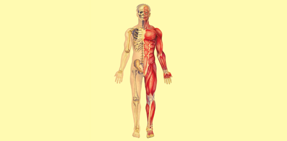
Human Anatomy, Physiology, and Kinesiology: A Student's Guide to Body Systems and Movement
Lesson Overview
- What Is the Relationship Between Anatomy, Physiology, and Kinesiology?
- How Do Organ Systems Cooperate to Maintain Homeostasis and Enable Activity?
- What Is the Structure and Function of Skeletal Muscles?
- How Is Muscle Contraction Controlled and What Is the Sliding Filament Theory?
- What Joints Exist in the Human Body and How Do They Influence Movement?
- How Does the Cardiovascular System Support Muscle Activity?
- What Are Common Pathologies Related to Anatomy and Movement?
- How Is the Nervous System Essential for Movement Coordination?
- What Are the Roles of the Endocrine and Urinary Systems in Physiology?
Many students and aspiring professionals struggle to connect how the body moves with why it moves that way. This lesson on human anatomy, physiology and kinesiology offers a structured way to understand these core principles. It simplifies complex systems into usable knowledge that supports academic success and real-world application in health sciences.
What Is the Relationship Between Anatomy, Physiology, and Kinesiology?
Many students face difficulty connecting the structure of the body to its function and movement. This section introduces how anatomy, physiology, and kinesiology interrelate, forming the foundation for understanding the human body in action.
- Anatomy defines the physical structure of organs, tissues, and systems.
- Physiology explains the biochemical and physical processes that keep these structures functioning.
- Kinesiology explores how muscles, bones, and joints produce coordinated movement.
These fields complement each other. Anatomy provides the blueprint, physiology offers the functional logic, and kinesiology applies both to dynamic motion and clinical application.
How Do Organ Systems Cooperate to Maintain Homeostasis and Enable Activity?
Each organ system works in conjunction with others to sustain homeostasis and enable function. This section explains the contribution of key systems to overall physiology, especially in relation to muscular function and movement.
- The skeletal system provides a rigid framework for attachment and protection.
- The muscular system enables voluntary and involuntary movements.
- The nervous system controls and coordinates activity via electrical impulses.
- The endocrine system modulates activity using chemical messengers.
- The cardiovascular and respiratory systems work together to deliver oxygen and remove waste products.
| Organ System | Main Functions | Key Interactions with Movement |
| Skeletal | Protection, mineral storage, leverage | Provides levers and joint structure |
| Muscular | Force production, heat generation | Initiates motion by contracting |
| Nervous | Sensory input, motor output | Transmits signals for muscle activation |
| Cardiovascular | Transport of gases, nutrients | Delivers oxygen to working muscles |
| Respiratory | Oxygen intake, CO2 removal | Supports aerobic metabolism in muscles |
| Endocrine | Hormonal control | Regulates metabolism and recovery |
What Is the Structure and Function of Skeletal Muscles?
Understanding skeletal muscle anatomy is essential for interpreting both movement and dysfunction. This section delves into muscle structure and physiological principles behind contraction.
- Skeletal muscle fibers are multinucleated cells made of repeating units called sarcomeres.
- Each sarcomere contains actin and myosin, the contractile proteins responsible for movement.
- The neuromuscular junction is where the nerve stimulates the muscle fiber.
Key Structural Elements:
| Component | Description | Function |
| Sarcomere | Basic unit of contraction | Shortens during contraction |
| Actin | Thin filament | Binding site for myosin |
| Myosin | Thick filament | Pulls actin to shorten sarcomere |
| Troponin-Tropomyosin | Regulatory proteins | Control access to actin |
Muscle Fiber Types:
| Fiber Type | Characteristics | Functional Role |
| Type I | Slow-twitch, high endurance | Posture, aerobic activities |
| Type IIa | Fast-twitch, moderate fatigue | Speed and strength activities |
| Type IIb | Fast-twitch, low endurance | High-intensity bursts |
How Is Muscle Contraction Controlled and What Is the Sliding Filament Theory?
Muscle contraction depends on neurological stimulation and biochemical interaction between filaments. This section explains the physiology behind voluntary muscle movement.
- A motor neuron sends an action potential to the neuromuscular junction.
- Acetylcholine (ACh) is released and binds to receptors on the sarcolemma.
- The action potential spreads through the T-tubules, triggering calcium release from the sarcoplasmic reticulum.
- Calcium binds to troponin, moving tropomyosin away from actin binding sites.
- Myosin heads attach, pull actin, and produce a contraction (cross-bridge cycling).
ATP is essential for both contraction and relaxation. Without ATP, myosin heads cannot detach, resulting in muscle rigidity (as seen in rigor mortis).
What Joints Exist in the Human Body and How Do They Influence Movement?
Joints determine the type and range of motion possible in the body. This section describes the structure, function, and classification of joints, emphasizing those most relevant to movement and kinesiology.
- Synovial joints are the most movable and include hinge, ball-and-socket, and saddle joints.
- Cartilaginous joints allow limited movement and are found in the spine.
- Fibrous joints are immovable and provide structural integrity (e.g., skull sutures).
Synovial Joint Characteristics:
| Joint Type | Examples | Movements |
| Hinge | Elbow, knee | Flexion, extension |
| Ball-and-socket | Shoulder, hip | Rotation, abduction, circumduction |
| Saddle | Thumb | Biaxial movement |
| Pivot | Atlas-axis | Rotation only |
Ligaments provide joint stability, while tendons attach muscles to bones, transmitting force to enable movement.
How Does the Cardiovascular System Support Muscle Activity?
The cardiovascular system maintains performance by supplying oxygen and nutrients and removing metabolic waste.
- The heart pumps deoxygenated blood to the lungs (pulmonary circulation) and oxygenated blood to the body (systemic circulation).
- During exercise, cardiac output increases due to higher heart rate and stroke volume.
- Blood is redirected from nonessential organs to active muscles.
Blood Composition:
| Component | Function |
| Red blood cells | Transport oxygen via hemoglobin |
| White blood cells | Immune defense |
| Platelets | Promote blood clotting |
| Plasma | Medium for transport |
Oxygen delivery is enhanced through increased breathing rate and deeper inhalations controlled by the respiratory centers in the brainstem.
What Are Common Pathologies Related to Anatomy and Movement?
Studying pathological conditions helps students understand deviations from normal function. This section covers key clinical examples that relate to the systems discussed.
- Myocardial infarction is caused by ischemia due to arterial blockage. It results in necrosis of cardiac tissue.
- Scoliosis involves a lateral spinal curvature, often identified during adolescence.
- Edema is the accumulation of interstitial fluid, commonly caused by heart failure or lymphatic dysfunction.
- Embolus refers to a traveling clot or air bubble that may obstruct a blood vessel.
- Plantar fasciitis and muscle strain are common musculoskeletal injuries linked to overuse or poor biomechanics.
Understanding these conditions improves clinical reasoning and diagnostic skills.
How Is the Nervous System Essential for Movement Coordination?
The nervous system controls voluntary and involuntary actions. This section highlights how electrical impulses generate motor responses and maintain equilibrium.
- The brain initiates voluntary movement through the motor cortex.
- The spinal cord relays messages to peripheral nerves.
- The cerebellum and basal ganglia modulate balance, coordination, and muscle tone.
- Sensory feedback from proprioceptors informs the CNS about joint position and muscle tension.
Motor Pathways:
| Structure | Function |
| Upper motor neurons | Originate in cortex; plan movements |
| Lower motor neurons | Exit spinal cord; activate muscles |
| Reflex arcs | Bypass brain for quick responses |
Disorders such as Parkinson's disease, multiple sclerosis, and peripheral neuropathy impair motor control and highlight the importance of neural integration.
What Are the Roles of the Endocrine and Urinary Systems in Physiology?
These systems help regulate internal balance and eliminate waste. Their integration supports homeostasis and performance.
- The adrenal glands release epinephrine and cortisol in response to stress.
- The pancreas regulates blood sugar by secreting insulin and glucagon.
- The kidneys filter blood, remove urea, and balance fluid and electrolytes.
The nephron is the kidney's functional unit. It performs filtration, reabsorption, secretion, and excretion of substances.
| Nephron Segment | Role |
| Glomerulus | Filters plasma |
| Proximal tubule | Reabsorbs nutrients, water |
| Loop of Henle | Concentrates urine |
| Distal tubule | Hormonal control |
| Collecting duct | Final urine composition |
Proper hydration, hormonal regulation, and pH balance are vital for muscle recovery and function.
Rate this lesson:
 Back to top

