Tissues and the Integumentary System: A Student’s Biology Guide
Tissues are groups of specialized cells that work together to perform specific functions. These are the building blocks of organs. The body's four primary tissue types-epithelial, connective, muscle, and nervous-combine in organs like the skin, which also forms the core of the integumentary system.
This system acts as the body's barrier, regulates temperature, and provides sensory input. Understanding how each tissue functions and contributes to the structure of the skin helps explain its protective and regulatory roles.
Epithelial Tissue
Epithelial tissue covers body surfaces, lines internal cavities and tubes, and forms glands. Epithelial cells are tightly packed with little extracellular material, forming continuous sheets.
These tissues serve as protective barriers and are often involved in absorption, secretion, and sensation. A defining feature of epithelia is polarity – cells have a top and bottom side with different structures and roles:
- Simple Cuboidal Epithelium: Found in glandular tissues; allows secretion and absorption.
- Apical Surface Function: The top of epithelial cells; faces the lumen.
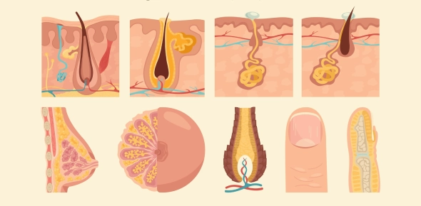
- Goblet Cells: Produce mucus in respiratory and digestive tracts. Dysfunction leads to mucus deficiency.
- Apocrine Secretion: Releases the product along with part of the cell's apical portion (e.g., mammary glands).
Connective Tissue
Connective tissues are the most abundant and widely distributed tissues in the body. They support, bind, and protect other tissues and organs.
Types and Functions:
| Type | Key Features | Location / Function |
| Cartilage | Avascular, lacks nerves, firm matrix | Ends of long bones, nose, trachea |
| Tendon | Dense regular CT, collagen-rich | Connects muscle to bone (e.g., Achilles tendon) |
| Reticular CT | Fibrous scaffold with reticular fibers | Supports lymph nodes, spleen, bone marrow |
| Blood (Plasma Matrix) | Liquid matrix; cells suspended in plasma | Transports gases, nutrients, and wastes |
| Adipose Tissue | Fat storage, insulation | Subcutaneous layer (hypodermis) |
- Achilles Tendon: Made of dense connective tissue; collagen provides strength.
- Blood Matrix: Not erythrocytes; the matrix is plasma.
- Ectodermal Origin: The nervous system derives from the ectoderm, affecting its development when disrupted.
(279).jpg)
Muscle Tissue
Muscle tissue is specialized for contraction and movement. Muscle cells (often called muscle fibers) contain contractile proteins (actin and myosin) that allow them to shorten (contract) and generate force.
There are three types of muscle tissue: skeletal muscle, cardiac muscle, and smooth muscle, each with distinct structure and function. A helpful way to organize this is to compare their key features:
Comparison Table:
| Type | Striations | Nuclei | Location | Control |
| Skeletal | Yes | Multinucleated | Attached to bones | Voluntary |
| Cardiac | Yes | Usually one | Heart wall | Involuntary |
| Smooth | No | One (central) | Walls of hollow organs | Involuntary |
- Smooth Muscle: Unstriated, uninucleated; controls movement in organs like the intestine and blood vessels.
- Limb Movement: Function of skeletal, not smooth muscle.
- Capillaries and Muscle: Smooth muscle controls blood vessel diameter (though true capillaries lack muscle in their walls).
Nervous Tissue
Nervous tissue is specialized for communication via electrical and chemical signals. It makes up the brain, spinal cord, and nerves. Nervous tissue allows the body to sense stimuli, process information, and transmit instructions. The primary cell type is the neuron, supported by various glial cells (neuroglia).
- Neurons: Transmit signals; respond to environmental changes.
- Functions: Accept stimuli and conduct electrical impulses (not involved in heat generation).
- Developmental Link: Derived from ectoderm; damage during development affects the nervous system.
Integumentary System
The integumentary system consists of the skin and its accessory structures (hair, nails, and glands). The skin is a vital organ that forms a barrier between our bodies and the external environment. It protects against injury, infection, and dehydration, helps regulate body temperature, and allows us to sense the world through touch.
Let's break down the components:
Skin Structure
| Layer | Key Features |
| Epidermis | Outer layer; stratified squamous epithelium; avascular |
| Dermis | Connective tissue; vascular, contains glands and nerves |
| Hypodermis | Subcutaneous fat; cushions, insulates, anchors skin |
- Stratum Corneum: First barrier a needle passes through; composed of dead keratinized cells.
- Epidermis and Touch: Merkel cells detect light touch; damage here can reduce sensation.
Epidermal Cell Types
- Keratinocytes: Main skin cells; produce keratin for protection.
- Melanocytes: Produce melanin; UV protection.
- Langerhans Cells: Immune defense.
- Merkel Cells: Sensory reception for light touch.
- Squamous Cell Carcinoma: Originates in keratinocytes of the stratum spinosum.
Dermal Layers
| Layer | Components |
| Papillary Layer | Areolar tissue, capillaries, Meissner's corpuscles |
| Reticular Layer | Dense irregular CT, houses glands, hair follicles |
- Sensory Receptors: Include Meissner's corpuscles (light touch), Pacinian corpuscles (deep pressure), and free nerve endings (pain, temperature).
Hypodermis
- Adipose Tissue: Stores fat, insulates, anchors skin.
- Injection Site: Hypodermic needles pass through epidermis to reach this layer.
Hair and Glands
- Hair: Provides light touch sensation and thermoregulation.
- Arrector Pili Muscle: Raises hair (goosebumps).
- Thermoregulation Role: Insulates by trapping heat.
Gland Types
| Gland | Secretion | Function / Location |
| Eccrine | Watery sweat | Cooling; found on palms, soles, forehead |
| Apocrine | Thick, lipid-rich sweat | Emotional/stress response; armpits, groin |
| Sebaceous | Sebum (oil) | Lubricates hair/skin; holocrine secretion |
- Eccrine Glands: Important for temperature regulation.
- Apocrine Glands: Begin at puberty; secrete into hair follicles.
- Sebaceous Glands: Use holocrine secretion; linked to acne.
(136).jpg)
Skin Functions
- Protection: Barrier against pathogens, UV, dehydration.
- Sensation: Nerve endings detect touch, pressure, temperature, and pain.
- Thermoregulation: Via sweat, blood flow, and hair.
- Excretion: Sweat removes waste.
- Vitamin D Synthesis: Triggered by UV exposure.
- Water Retention: Lipid layers prevent fluid loss.
Rate this lesson:
 Back to top