Assessment Of The Cat Appendicular Skeleton

Approved & Edited by ProProfs Editorial Team
The editorial team at ProProfs Quizzes consists of a select group of subject experts, trivia writers, and quiz masters who have authored over 10,000 quizzes taken by more than 100 million users. This team includes our in-house seasoned quiz moderators and subject matter experts. Our editorial experts, spread across the world, are rigorously trained using our comprehensive guidelines to ensure that you receive the highest quality quizzes.
Learn about Our Editorial Process
| By Landonbrewer
L
Landonbrewer
Community Contributor
Quizzes Created: 1 | Total Attempts: 1,768
Questions: 58 | Attempts: 1,770

SettingsSettingsSettings
Assessment Of The Cat Appendicular Skeleton - Quiz

This assessment consists only of structure identification of the cat appendicual skeleton.


Questions and Answers
  • 1. 

    What is structure 2?

    Explanation
    The supraspinous fossa is a specific structure in the human body. It is a shallow depression located on the posterior side of the scapula, specifically above the spine of the scapula. This fossa serves as an attachment site for muscles and ligaments, including the supraspinatus muscle. It is important for the movement and stability of the shoulder joint.

    Rate this question:

  • 2. 

    Name structure 3

    Explanation
    The given answer, "scapular spine," is correct because it accurately identifies the structure being described. The scapular spine is a prominent ridge of bone on the posterior surface of the scapula (shoulder blade). It runs diagonally across the scapula and provides attachment for several muscles, including the deltoid and trapezius. The scapular spine helps to provide stability and support to the shoulder joint, allowing for the movement and rotation of the arm.

    Rate this question:

  • 3. 

    Name structure 4

    Explanation
    The infraspinous fossa is a name structure located on the posterior surface of the scapula bone. It is a concave depression that is found below the spine of the scapula. The term "infraspinous" refers to its position below the spine, and "fossa" indicates that it is a shallow, hollowed-out area. This anatomical feature serves as an attachment site for muscles and ligaments, contributing to the movement and stability of the shoulder joint.

    Rate this question:

  • 4. 

    What is structure 5?

  • 5. 

    What is structure 6?

    Explanation
    Structure 6 refers to the acromion. The acromion is a bony process on the scapula (shoulder blade) that forms the highest point of the shoulder. It is easily identifiable as a prominent, triangular-shaped projection. The acromion plays a crucial role in the stability and movement of the shoulder joint, as it provides attachment points for various muscles and ligaments.

    Rate this question:

  • 6. 

    What is structure 7?

    Explanation
    Structure 7 refers to the subscapular fossa. The subscapular fossa is a concave depression located on the anterior surface of the scapula. It is the largest of the three fossae on the scapula, with the other two being the supraspinous fossa and infraspinous fossa. The subscapular fossa provides attachment for the subscapularis muscle, which is responsible for internal rotation of the arm.

    Rate this question:

  • 7. 

    What is structure 8?

  • 8. 

    What is structure 9?

    Explanation
    The coracoid process is a structure located in the shoulder region. It is a hook-like projection that extends from the scapula bone. The coracoid process serves as an attachment point for various muscles and ligaments, providing stability and support to the shoulder joint.

    Rate this question:

  • 9. 

    What is structure 10?

    Explanation
    The correct answer for the question "What is structure 10?" is the posterior border.

    Rate this question:

  • 10. 

    What is structure 1?

    Explanation
    The lesser tuberosity is a bony prominence located on the anterior surface of the proximal end of the humerus bone. It serves as an attachment site for the subscapularis muscle, which is one of the four muscles that make up the rotator cuff. The lesser tuberosity helps stabilize and move the shoulder joint, allowing for various arm movements.

    Rate this question:

  • 11. 

    What is structure 2?

    Explanation
    The bicipital groove is a anatomical structure located on the humerus bone. It is a shallow groove that runs along the anterior surface of the humerus and serves as a pathway for the long head of the biceps tendon. This groove provides stability and protection for the tendon as it moves during arm movements. The bicipital groove is an important landmark in the shoulder region and is commonly used as a reference point in clinical examinations and surgical procedures.

    Rate this question:

  • 12. 

    What is structure 3?

    Explanation
    Structure 3 refers to a shaft.

    Rate this question:

  • 13. 

    What is structure 4?

    Explanation
    Structure 4 refers to the supracondyloid foramen. The supracondyloid foramen is an anatomical variation located above the medial epicondyle of the humerus. It is a small opening in the bone that allows for the passage of nerves and blood vessels. This anatomical feature is not present in all individuals, but when it is present, it can be an important landmark for surgeons and anatomists.

    Rate this question:

  • 14. 

    What is structure 5?

    Explanation
    Structure 5 refers to the greater tuberosity. The greater tuberosity is a bony prominence located on the proximal end of the humerus bone. It serves as an attachment site for various muscles and tendons, including the supraspinatus, infraspinatus, and teres minor muscles. The greater tuberosity plays an important role in shoulder movements and stability.

    Rate this question:

  • 15. 

    What is structure 6?

    Explanation
    Structure 6 refers to the head.

    Rate this question:

  • 16. 

    What is structure 7?

    Explanation
    Structure 7 refers to the lateral epicondyle. The lateral epicondyle is a bony prominence located on the outer side of the humerus bone in the upper arm. It serves as an attachment point for various muscles and ligaments, including those involved in the movement of the forearm and wrist. The lateral epicondyle is commonly associated with conditions such as tennis elbow, which is characterized by pain and inflammation in the area.

    Rate this question:

  • 17. 

    What is structure 8?

    Explanation
    The structure 8 referred to in the question is the olecranon fossa. The olecranon fossa is a depression located on the posterior side of the humerus bone, specifically in the distal end of the humerus. It is a concave area that accommodates the olecranon process of the ulna bone during extension of the forearm. This allows for smooth movement and flexibility of the elbow joint.

    Rate this question:

  • 18. 

    What is structure 9?

    Explanation
    Structure 9 refers to the medial epicondyle. The medial epicondyle is a bony prominence located on the inner side of the humerus bone in the upper arm. It serves as an attachment point for various muscles and ligaments, including the flexor muscles of the forearm. This structure can be easily palpated and is important in understanding the anatomy and function of the elbow joint.

    Rate this question:

  • 19. 

    What is structure 1?

    Explanation
    The structure 1 is referred to as the olecranon.

    Rate this question:

  • 20. 

    What is structure 2?

    Explanation
    The trochlear notch is a feature found in the structure 2. It is a depression or groove located on the ulna bone, which is one of the two bones in the forearm. The trochlear notch articulates with the trochlea of the humerus bone to form the elbow joint. This allows for the flexion and extension of the forearm.

    Rate this question:

  • 21. 

    What is structure 3?

    Explanation
    The coronoid process is a bony projection located on the anterior surface of the ulna bone. It is part of the ulna bone's structure and serves as an attachment point for muscles involved in forearm movement. The coronoid process acts as a stabilizer and helps prevent dislocation of the elbow joint.

    Rate this question:

  • 22. 

    What is structure 4?

    Explanation
    Structure 4 refers to the styloid process of the ulna. The styloid process is a bony projection located at the distal end of the ulna bone in the forearm. It serves as an attachment point for ligaments and tendons, providing stability and support to the wrist joint. The styloid process of the ulna can be palpated on the medial side of the wrist and is important for proper wrist movement and function.

    Rate this question:

  • 23. 

    What is structure 5?

  • 24. 

    What is structure 6?

  • 25. 

    What is structure 7?

    Explanation
    Structure 7 refers to the bicipital tuberosity. The bicipital tuberosity is a bony prominence located on the upper end of the radius bone in the forearm. It serves as the attachment point for the biceps brachii muscle, which is responsible for flexing the elbow joint. The bicipital tuberosity is easily palpable and can be used as a landmark for locating the insertion of the biceps brachii tendon.

    Rate this question:

  • 26. 

    What is structure 8?

    Explanation
    Structure 8 refers to the interosseous crest of the radius. The interosseous crest is a ridge-like structure located on the medial side of the radius bone. It serves as an attachment site for various muscles and ligaments, providing stability and support to the forearm.

    Rate this question:

  • 27. 

    What is structure 9?

    Explanation
    Structure 9 refers to the styloid process of the radius. The styloid process is a bony projection located at the distal end of the radius bone in the forearm. It serves as an attachment point for ligaments and muscles, providing stability and support to the wrist joint. The styloid process of the radius can be palpated on the lateral side of the wrist and is an important landmark for anatomical reference.

    Rate this question:

  • 28. 

    What is structure 6?

    Explanation
    Structure 6 refers to carpals, which are a group of small bones located in the wrist. These bones are arranged in two rows and play a crucial role in providing stability and flexibility to the wrist joint. Carpals are responsible for various movements of the hand and are essential for performing fine motor skills.

    Rate this question:

  • 29. 

    What is structure 7?

  • 30. 

    What is structure 8?

    Explanation
    Structure 8 refers to the phalanges. Phalanges are the bones that make up the fingers and toes. They are long, slender bones that articulate with the metacarpals in the hands and the metatarsals in the feet. The phalanges play a crucial role in enabling fine motor movements and providing support for the hands and feet.

    Rate this question:

  • 31. 

    Does this picture suggest that this animal is a plantigrade, digitigrade, or unguligrade?

    Explanation
    This picture suggests that the animal in question is a digitigrade. Digitigrade refers to animals that walk on their toes, with their heel elevated off the ground. This can be inferred from the posture and positioning of the animal's legs in the picture, indicating that it walks on its toes rather than the entire foot or hooves.

    Rate this question:

  • 32. 

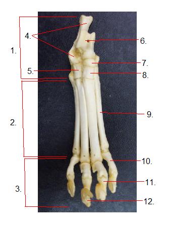
    What is structure 1?

    Explanation
    The structure 1 referred to in the question is tarsals. Tarsals are a group of seven bones located in the foot region. They are responsible for providing stability and support to the foot, allowing for movement and balance. These bones are arranged in a specific pattern to form the structure of the foot.

    Rate this question:

  • 33. 

    What is structure 2?

    Explanation
    Structure 2 refers to the metatarsals. The metatarsals are a group of long bones located in the foot, connecting the tarsal bones to the phalanges. They are responsible for providing support and stability to the foot, as well as playing a role in weight-bearing and walking.

    Rate this question:

  • 34. 

    What is structure 3?

    Explanation
    Structure 3 refers to phalanges. The phalanges are the bones that make up the fingers and toes. They are long, slender bones that are connected to the metacarpals in the hands and the metatarsals in the feet. The phalanges play a crucial role in allowing for fine motor movements and providing support and flexibility to the hands and feet.

    Rate this question:

  • 35. 

    What is structure 4?

    Explanation
    The structure referred to as "structure 4" is the calcaneus.

    Rate this question:

  • 36. 

    What is structure 1?

    Explanation
    The iliac crest is a prominent ridge of bone located on the top of the hip bone. It forms the upper border of the pelvis and can be felt and seen on the side of the body. The iliac crest serves as an attachment site for several muscles and ligaments, including the abdominal muscles and the iliotibial band. It also provides support and stability to the pelvis and helps protect the internal organs in the abdominal cavity.

    Rate this question:

  • 37. 

    What is structure 2?

  • 38. 

    What is structure 3?

    Explanation
    Structure 3 is the ilium, which is one of the three bones that make up the hip bone. The ilium is the largest and uppermost bone of the hip, forming the prominence of the hip. It helps to support the weight of the upper body and provides attachment points for various muscles and ligaments.

    Rate this question:

  • 39. 

    What is structure 4?

    Explanation
    Structure 4 refers to the acetabulum. The acetabulum is a part of the hip bone and is a socket-like structure that forms the hip joint. It is located on the lateral side of the pelvis and serves as the point of articulation for the head of the femur (thigh bone). The acetabulum is responsible for providing stability and support to the hip joint, allowing for smooth movement and weight-bearing activities.

    Rate this question:

  • 40. 

    What is structure 5?

    Explanation
    Structure 5 refers to the pubis.

    Rate this question:

  • 41. 

    What is structure 6?

    Explanation
    The structure 6 refers to the obturator foramen. The obturator foramen is a large, oval-shaped opening in the hip bone, formed by the ischium and pubis bones. It is located in the pelvic region and serves as a passageway for blood vessels, nerves, and muscles that supply the lower limb.

    Rate this question:

  • 42. 

    What is structure 7?

    Explanation
    Structure 7 refers to the ischium. The ischium is one of the three bones that make up the hip bone, along with the ilium and pubis. It is located at the lower and posterior part of the hip bone and forms the posterior portion of the pelvis. The ischium is commonly known as the "sit bone" as it bears the weight when sitting. It also provides attachment points for various muscles and ligaments in the pelvic region.

    Rate this question:

  • 43. 

    What is structure 8?

    Explanation
    Structure 8 refers to the wing of the Ilium. The Ilium is one of the bones that make up the pelvis, and it has a wing-like structure that extends outwards. This wing of the Ilium provides attachment points for muscles and ligaments, and it helps to support and stabilize the pelvis.

    Rate this question:

  • 44. 

    What is structure 10?

    Explanation
    Structure 10 refers to the ischial tuberosity. The ischial tuberosity is a bony prominence located at the base of the pelvis, specifically on the ischium bone. It is commonly known as the "sitting bone" as it bears the weight when a person is seated. The ischial tuberosity is important for providing support and stability to the pelvis and is involved in various functions such as sitting, walking, and running.

    Rate this question:

  • 45. 

    What is structure 11?

    Explanation
    Structure 11 refers to the pelvic symphysis. The pelvic symphysis is a joint located between the two pubic bones in the pelvis. It is a cartilaginous joint that connects the left and right pubic bones, allowing them to move slightly during childbirth. The pelvic symphysis also provides stability and support to the pelvis.

    Rate this question:

  • 46. 

    What is structure 1?

  • 47. 

    What is structure 2?

    Explanation
    Structure 2 refers to the neck.

    Rate this question:

  • 48. 

    What is structure 3?

    Explanation
    Structure 3 refers to the greater trochanter. The greater trochanter is a bony prominence located on the femur, specifically on the proximal and lateral side. It serves as a site for muscle attachment, particularly for the gluteus medius and gluteus minimus muscles. The greater trochanter plays a crucial role in hip movement, stability, and overall function.

    Rate this question:

  • 49. 

    What is structure 4?

  • 50. 

    What is structure 5?

    Explanation
    Structure 5 refers to the patellar trochlea. The patellar trochlea is a groove on the femur bone that allows the patella (kneecap) to move smoothly during knee flexion and extension. It serves as a track for the patella to slide up and down, providing stability and facilitating proper movement of the knee joint.

    Rate this question:

Quiz Review Timeline +

Our quizzes are rigorously reviewed, monitored and continuously updated by our expert board to maintain accuracy, relevance, and timeliness.

  • Current Version
  • Mar 20, 2023
    Quiz Edited by
    ProProfs Editorial Team
  • Apr 24, 2009
    Quiz Created by
    Landonbrewer
Back to Top Back to top
Advertisement
×

Wait!
Here's an interesting quiz for you.

We have other quizzes matching your interest.