Unit II: Overview Of The Skeleton & Appendicular Skeleton

Approved & Edited by ProProfs Editorial Team
The editorial team at ProProfs Quizzes consists of a select group of subject experts, trivia writers, and quiz masters who have authored over 10,000 quizzes taken by more than 100 million users. This team includes our in-house seasoned quiz moderators and subject matter experts. Our editorial experts, spread across the world, are rigorously trained using our comprehensive guidelines to ensure that you receive the highest quality quizzes.
Learn about Our Editorial Process
| By Sutton_anatomy
S
Sutton_anatomy
Community Contributor
Quizzes Created: 8 | Total Attempts: 27,715
Questions: 26 | Attempts: 880

SettingsSettingsSettings
Unit II: Overview Of The Skeleton & Appendicular Skeleton - Quiz

Questions and Answers
  • 1. 

    Identify bone marking A:

    Explanation
    The bone marking A is referred to as the acromial end. The term "acromial" is used to describe the part of the bone that is related to the acromion process, which is a bony projection on the scapula (shoulder blade). The acromial end is the part of the bone that articulates with the acromion process, forming a joint.

    Rate this question:

  • 2. 

    Identify bone marking B:

    Explanation
    The correct answer is Acromion, acromion. The bone marking B is the acromion. The acromion is a bony process on the scapula (shoulder blade) that forms the highest point of the shoulder. It is easily identified as a prominent, curved projection that extends laterally from the scapula. The term "acromion" refers to this specific bone marking on the scapula.

    Rate this question:

  • 3. 

    Identify bone marking C:

    Explanation
    The correct answer is supraspinous fossa. The supraspinous fossa is a bone marking located on the scapula (shoulder blade). It is a shallow depression on the posterior side of the scapula, above the spine of the scapula. It serves as an attachment site for muscles and ligaments involved in shoulder movement and stability.

    Rate this question:

  • 4. 

    Identify bone marking A:

    Explanation
    The correct answer is the Medial Epicondyle. The term "medial" refers to the side of the body that is closer to the midline, while "epicondyle" refers to a bony prominence located above a condyle. Therefore, the Medial Epicondyle is a bony prominence on the inner side of the elbow joint.

    Rate this question:

  • 5. 

    Identify bone marking B:

    Explanation
    The bone marking B is called the coronoid fossa.

    Rate this question:

  • 6. 

    Identify bone marking C:

    Explanation
    The bone marking C is called the capitulum. The repetition of the term "capitulum" in the answer suggests that it is the correct option.

    Rate this question:

  • 7. 

    Is bone A medial or lateral?

    Explanation
    The bone A is described as lateral. This means that it is positioned on the outer side or away from the midline of the body.

    Rate this question:

  • 8. 

    Identify bone marking A:

    Explanation
    The correct answer is "Radial Tuberosity". The radial tuberosity is a bone marking located on the radius bone of the forearm. It is a rough, bumpy area where the biceps muscle attaches. The repetition of the term "Radial Tuberosity" in different formats indicates that it is the correct answer.

    Rate this question:

  • 9. 

    Identify bone marking A:

    Explanation
    The correct answer is "Trochlear Notch". The trochlear notch is a V-shaped depression located on the proximal end of the ulna bone. It articulates with the trochlea of the humerus to form the elbow joint. This notch allows for the smooth movement of the ulna during flexion and extension of the forearm.

    Rate this question:

  • 10. 

    Identify bone marking B:

    Explanation
    The correct answer is "Radial Notch, Radial notch, radial notch." The bone marking being referred to in this question is the radial notch. It is a small depression or indentation on the radius bone, located near the distal end. This marking provides a space for the ulna bone to articulate with the radius bone, allowing for movement and rotation of the forearm. The repetition of the term "radial notch" in different capitalizations suggests that it is the correct answer.

    Rate this question:

  • 11. 

    Identify bone A:

    Explanation
    The bone A is identified as the distal phalange.

    Rate this question:

  • 12. 

    Identify bone marking A:

    Explanation
    The correct answer is "Anterior Inferior Iliac Spine", "Anterior inferior iliac spine", and "anterior inferior iliac spine". These are all correct because they refer to the same bone marking. The anterior inferior iliac spine is a bony projection located on the anterior and inferior aspect of the ilium bone in the pelvis. It serves as an attachment point for various muscles and ligaments.

    Rate this question:

  • 13. 

    Identify bone marking C:

    Explanation
    The correct answer is "Ischial Spine, Ischial spine, ischial spine." The ischial spine is a bone marking located on the ischium, which is part of the pelvis. It is a small projection that extends posteriorly and superiorly.

    Rate this question:

  • 14. 

    The bone shown below represents the os coxa.

    • A.

      True

    • B.

      False

    Correct Answer
    A. True
    Explanation
    The bone shown below represents the os coxa. The os coxa, also known as the hip bone, is a large flat bone located on each side of the pelvis. It consists of three fused bones: the ilium, ischium, and pubis. These bones come together to form a socket called the acetabulum, which connects the hip joint and the femur. Therefore, the statement "The bone shown below represents the os coxa" is true.

    Rate this question:

  • 15. 

    Identify bone marking A:

    Correct Answer
    Greater Sciatic Notch
    Greater sciatic notch
    greater sciatic notch
    Explanation
    The correct answer is "Greater Sciatic Notch". The greater sciatic notch is a large indentation on the posterior aspect of the hip bone. It is located below the posterior inferior iliac spine and above the ischial spine. The greater sciatic notch allows for the passage of the sciatic nerve, as well as blood vessels and other structures, between the pelvis and the lower limb.

    Rate this question:

  • 16. 

    Identify bone marking A:

    Correct Answer
    Neck
    neck
  • 17. 

    Identify bone marking A:

    Correct Answer
    Intercondylar Fossa
    Intercondylar fossa
    intercondylar fossa
    Explanation
    The correct answer is "Intercondylar Fossa, Intercondylar fossa, intercondylar fossa." This bone marking refers to a depression located between the condyles of a bone, typically found in the knee joint area. It allows for the articulation and movement of the bones in the joint.

    Rate this question:

  • 18. 

    Identify bone marking A:

    Correct Answer
    Lateral Condyle
    Lateral condyle
    lateral condyle
    Explanation
    The bone marking A is the lateral condyle. The term "lateral condyle" refers to a rounded prominence on the lateral side of a bone, typically found at a joint. In this case, it is specifically referring to the lateral condyle of a bone.

    Rate this question:

  • 19. 

    The bone shown below represents the tibia.

    • A.

      True

    • B.

      False

    Correct Answer
    B. False
    Explanation
    The given statement is false because the bone shown below does not represent the tibia.

    Rate this question:

  • 20. 

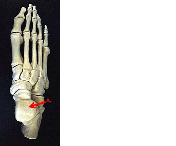
    Identify bone A:

    Correct Answer
    Talus
    talus
    Explanation
    The bone that is identified as "A" is called the talus. The talus is a bone located in the foot, specifically in the ankle joint. It is an important bone that connects the leg bones (tibia and fibula) to the foot bones. The talus plays a crucial role in allowing movement and flexibility of the foot and ankle.

    Rate this question:

  • 21. 

    Identify A:

    Correct Answer
    Articular Cartilage
    Articular cartilage
    articular cartilage
    Explanation
    The correct answer for the given question is "Articular Cartilage, Articular cartilage, articular cartilage". This suggests that all three options are correct and refer to the same thing, which is the articular cartilage. The repetition of the term in different capitalization styles may be due to formatting or typing errors, but they all point to the same anatomical structure.

    Rate this question:

  • 22. 

    Identify B:

    Correct Answer
    Endosteum
    endosteum
    Explanation
    The correct answer is "Endosteum, endosteum." The endosteum is a thin layer of connective tissue that lines the inner surface of the bone. It contains osteoprogenitor cells, which are responsible for bone growth and repair. The repetition of "endosteum" in the answer suggests that B refers to the endosteum.

    Rate this question:

  • 23. 

    Trochanters are found only on the femur.

    • A.

      True

    • B.

      False

    Correct Answer
    A. True
    Explanation
    Trochanters are bony protrusions found on the femur bone. They serve as attachment points for muscles, tendons, and ligaments, providing stability and facilitating movement of the hip joint. The trochanters include the greater trochanter and the lesser trochanter, which are located at specific points on the femur. Therefore, it is true that trochanters are found only on the femur bone.

    Rate this question:

  • 24. 

    Teeth are actually part of the digestive system not the skeletal system despite being composed of largely bone.

    • A.

      True

    • B.

      False

    Correct Answer
    B. False
    Explanation
    Teeth are not part of the digestive system; they are actually part of the skeletal system. Although teeth are composed largely of bone, they are not involved in the actual digestion of food. Instead, teeth play a crucial role in the initial mechanical breakdown of food, aiding in the process of chewing and grinding before it enters the digestive system.

    Rate this question:

  • 25. 

    In a male pelvis the ischial spines flare inward.

    • A.

      True

    • B.

      False

    Correct Answer
    A. True
    Explanation
    In a male pelvis, the ischial spines do indeed flare inward. This anatomical feature is one of the key differences between male and female pelves. The flaring of the ischial spines in males allows for a narrower pelvic outlet, which is advantageous for supporting the weight of the upper body and facilitating bipedal locomotion. In contrast, the ischial spines in a female pelvis are wider apart, providing a larger pelvic outlet to accommodate childbirth.

    Rate this question:

  • 26. 

    Which of these bones forms within a tendon?

    • A.

      Os Coxae

    • B.

      Patella

    • C.

      Carpals

    • D.

      Clavicle

    Correct Answer
    B. Patella
    Explanation
    The patella, commonly known as the kneecap, is a sesamoid bone that forms within the tendon of the quadriceps muscle. It is located in front of the knee joint and helps to protect the joint and provide mechanical advantage to the muscles involved in knee extension. The other options, including Os Coxae (hip bone), Carpals (wrist bones), and Clavicle (collarbone), do not form within a tendon.

    Rate this question:

Quiz Review Timeline +

Our quizzes are rigorously reviewed, monitored and continuously updated by our expert board to maintain accuracy, relevance, and timeliness.

  • Current Version
  • Mar 21, 2023
    Quiz Edited by
    ProProfs Editorial Team
  • Jan 07, 2012
    Quiz Created by
    Sutton_anatomy
Back to Top Back to top
Advertisement
×

Wait!
Here's an interesting quiz for you.

We have other quizzes matching your interest.