What Is Anatomy and Physiology of Sensory Organs Structure & Function?
Lesson Overview
- What Are Sensory Organs and Why Are They Vital to Human Survival?
- How Does the Human Eye Convert Light into Visual Signals?
- How Does the Ear Detect Sound Waves and Maintain Balance?
- How Does the Nose Detect Odor Molecules?
- How Does the Tongue Detect and Interpret Different Tastes?
- How Does the Skin Function as a Sensory Organ?
- What Are the Common Disorders Associated With Sensory Organs?
- How Are Sensory Functions Tested in Clinical Settings?
- Conclusion
When Sam couldn't explain the difference between rods and cones in his biology oral test, he realized just reading labels on diagrams wasn't enough. Understanding the anatomy and physiology of the sensory organs means learning how sight, sound, touch, taste, and smell work together. This lesson makes it easy and complete.
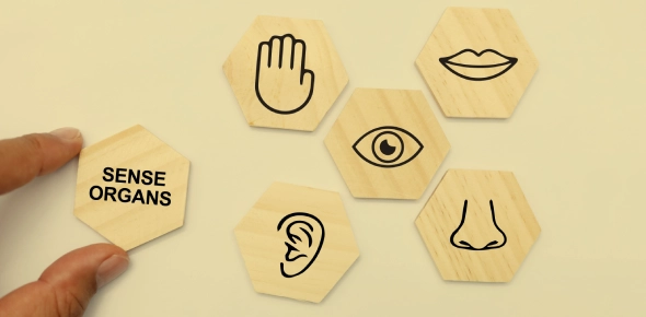
What Are Sensory Organs and Why Are They Vital to Human Survival?
This section introduces sensory organs as essential biological structures that allow organisms to perceive and respond to their environment.
Sensory organs are specialized anatomical systems designed to detect and transmit environmental information to the central nervous system. Each sensory organ responds to a specific type of stimulus: light, sound, chemical molecules, pressure, or temperature. The integration of sensory input helps organisms to make decisions, coordinate movements, and ensure safety and survival. The five major senses-vision, hearing, smell, taste, and touch-are mediated by the eyes, ears, nose, tongue, and skin respectively.
Major Roles of Sensory Organs:
- Convert external stimuli into electrical impulses (transduction)
- Send sensory data to the brain for interpretation
- Maintain awareness of internal and external environments
- Contribute to memory, learning, and emotional responses
How Does the Human Eye Convert Light into Visual Signals?
This section explores the complex anatomy of the eye and the physiological processes involved in visual perception.
The human eye functions as a highly specialized organ for photoreception. Light enters the eye through the cornea and passes through the aqueous humor, pupil, and lens before reaching the retina.
Anatomical Features of the Eye:
- Cornea: Transparent anterior layer that refracts light onto the lens
- Pupil and Iris: Regulate the amount of light entering the eye
- Lens: Biconvex structure that changes shape for near or distant focus (accommodation)
- Retina: Neural layer containing photoreceptors (rods and cones)
- Optic Nerve: Transmits electrical impulses to the brain
Photoreceptor Cells:
- Rods: Enable scotopic (low light) vision and detect shades of gray
- Cones: Enable photopic (bright light) vision and detect red, green, and blue wavelengths
Macula and Fovea:
- The macula is the region of central vision.
- The fovea within the macula provides the highest acuity and concentration of cones.
Photoreceptors and Their Functions
| Cell Type | Function | Light Sensitivity | Color Detection | Location |
| Rods | Dim light vision | High | No | Peripheral retina |
| Cones | Color and detail vision | Moderate | Yes (RGB) | Central retina |
How Does the Ear Detect Sound Waves and Maintain Balance?
This section explains the ear's anatomy and function in auditory transduction and equilibrium.
The ear is divided into three regions: external ear, middle ear, and inner ear. Each section performs distinct roles in sound perception and balance.
External Ear:
- Auricle (Pinna): Collects sound waves
- External Auditory Canal: Channels sound to the tympanic membrane
Middle Ear:
- Tympanic Membrane (Eardrum): Vibrates in response to sound
- Ossicles (Malleus, Incus, Stapes): Transmit and amplify vibrations
- Eustachian Tube: Connects to pharynx to equalize air pressure
Inner Ear:
- Cochlea: Converts mechanical vibrations into neural signals
- Organ of Corti: Contains hair cells responsible for auditory transduction
- Vestibule and Semicircular Canals: Detect gravity and rotational movement for balance
Inner Ear Components and Functions
| Structure | Role |
| Cochlea | Auditory perception |
| Semicircular Canals | Rotational motion detection |
| Vestibule | Linear acceleration and gravity |
| Oval Window | Entry point for stapes vibrations |
| Perilymph | Fluid that transmits vibrations |
How Does the Nose Detect Odor Molecules?
This section outlines the olfactory system's mechanism for detecting airborne chemical stimuli.
The sense of smell, or olfaction, begins in the olfactory epithelium located in the upper nasal cavity. This region contains receptor cells, support cells, and basal cells.
Key Olfactory Components:
- Olfactory Hairs: Cilia that bind odorant molecules
- Receptor Cells: Neurons that generate action potentials
- Cribriform Plate: Bone through which olfactory axons pass
- Olfactory Bulb: Brain structure that processes odor signals
Signals travel to the olfactory cortex, limbic system, and hypothalamus, influencing emotion and behavior.
Olfactory Pathway Overview
| Step | Action |
| Odorant binds | Olfactory receptor in nasal cavity |
| Signal sent | Through olfactory nerve to bulb |
| Processed in | Olfactory cortex and limbic system |
How Does the Tongue Detect and Interpret Different Tastes?
This section explores gustatory anatomy and the chemical basis of taste perception.
Taste is detected by taste buds located on various types of lingual papillae. Each taste bud contains gustatory cells with microvilli that detect dissolved chemicals.
Taste Modalities:
- Sweet: Detected at the tip of the tongue; signals sugars
- Salty: Detected along the lateral margins; signals electrolytes
- Sour: Signals acidity (hydrogen ions)
- Bitter: Detects alkaloids, often toxic
- Umami: Detects amino acids like glutamate
Transmission:
- CN VII (Facial) – Anterior 2/3 of tongue
- CN IX (Glossopharyngeal) – Posterior 1/3
- CN X (Vagus) – Pharynx and epiglottis
Table: Taste and Their Functions
| Taste | Chemical Stimulus | Evolutionary Function |
| Sweet | Glucose, fructose | Energy source |
| Salty | Sodium chloride | Electrolyte balance |
| Sour | Acids | pH detection |
| Bitter | Alkaloids | Toxin warning |
| Umami | Glutamate, nucleotides | Protein recognition |
How Does the Skin Function as a Sensory Organ?
This section explains the cutaneous receptors responsible for tactile sensations.
The skin houses mechanoreceptors, thermoreceptors, and nociceptors that detect mechanical, temperature, and pain stimuli respectively.
Types of Touch Receptors:
- Meissner's Corpuscles: Detect fine touch and vibration
- Pacinian Corpuscles: Sense deep pressure and rapid vibrations
- Merkel Discs: Detect sustained pressure and texture
- Free Nerve Endings: Respond to temperature and pain
Distribution:
- Receptor density varies by body region (e.g., fingertips are most sensitive)
- Signals are relayed to the brain via dorsal spinal roots and cranial nerves
Table: Skin Receptor Summary
| Receptor Type | Detected Stimulus | Location |
| Meissner's corpuscles | Light touch | Dermal papillae (fingertips) |
| Pacinian corpuscles | Deep pressure | Deep dermis and joints |
| Merkel cells | Constant touch/pressure | Epidermal-dermal junction |
| Free nerve endings | Pain, temperature | Throughout skin |
What Are the Common Disorders Associated With Sensory Organs?
This section highlights disorders affecting vision, hearing, balance, and other sensory modalities.
Visual Disorders:
- Cataracts: Lens opacity impairs light transmission
- Macular Degeneration: Central retina degradation reduces sharp vision
- Achromatopsia: Genetic disorder resulting in total color blindness
- Retinoblastoma: Malignant retinal tumor, typically in children
Auditory and Vestibular Disorders:
- Otitis Media/Externa: Infection of middle/outer ear
- Tinnitus: Perception of phantom sounds (ringing)
- Vertigo: Balance disturbance due to inner ear issues
- Meniere's Disease: Inner ear disorder causing fluctuating hearing loss and dizziness
Other Sensory Conditions:
- Amblyopia: Visual development disorder leading to lazy eye
- Strabismus: Misalignment of the eyes
- Nyctalopia: Difficulty seeing in dim light (night blindness)
- Otopyorrhea: Ear infection with pus discharge
Table: Sensory Disorders Overview
| Disorder | Affected Organ | Key Symptoms |
| Cataracts | Eye | Blurry vision |
| Tinnitus | Ear | Ringing sounds |
| Vertigo | Inner Ear | Spinning sensation |
| Achromatopsia | Eye | Complete color blindness |
| Amblyopia | Eye | Reduced vision in one eye |
How Are Sensory Functions Tested in Clinical Settings?
This section describes diagnostic tools and their applications in evaluating sensory health.
Vision Testing:
- Snellen Chart: Assesses visual acuity (e.g., 20/20 vision)
- Ophthalmoscopy: Examines retina and optic nerve
Hearing Evaluation:
- Audiometry: Measures hearing sensitivity across frequencies
- Tuning Fork Tests: Identify conductive vs. sensorineural loss
Balance Testing:
- Romberg Test: Evaluates postural control and vestibular function
Taste and Smell Assessment:
- Taste Strip Testing: Uses calibrated stimuli to assess taste thresholds
- Olfactory Testing: Uses aromatic compounds to detect olfactory dysfunction
Tactile Sensitivity:
- Monofilament Testing: Evaluates pressure perception
- Two-point Discrimination: Assesses spatial touch resolution
Conclusion
This lesson provided an in-depth look at the anatomy and physiology of the sensory organs, including the eyes, ears, nose, tongue, and skin. Each organ contributes vital data to the central nervous system for interpretation and response. Understanding how these systems work together-and how disorders affect them-gives students a solid foundation for clinical, biological, and health science studies.
Take This Quiz
Rate this lesson:
 Back to top