Brain Anatomy: Structure, Primary Functions, and Neural Pathways Explained
Lesson Overview
The human brain is a complex organ divided into several major regions – primarily the cerebrum, the cerebellum, and the brainstem. Each of these parts has unique structures and functions. The cerebrum (the largest portion) handles higher cognitive functions; the cerebellum coordinates movement and balance; and the brainstem connects the brain to the spinal cord while controlling basic life functions.
The surface of the brain is highly folded into gyri (ridges) and sulci (grooves), which increase surface area for neurons. In this lesson, you will explore brain anatomy, covering the cerebral hemispheres and lobes, the brainstem (midbrain, pons, medulla), the cerebellum, major neural pathways, and the distinction between gray and white matter.
The Cerebrum and Cerebral Hemispheres
The cerebrum is the largest brain part, handling conscious thought, reasoning, sensory processing, and voluntary movement. It's split into left and right hemispheres by the longitudinal fissure. These halves communicate via the corpus callosum, a major nerve fiber bundle.
Signals from each body side cross over in the brainstem-a process called decussation-so the left brain controls the right body side and vice versa. The outer cerebrum, the cortex, is rich in neurons and organized into folds: gyri (ridges) and sulci (grooves). These increase surface area for neural activity.
Cerebral Lobes and Their Functions
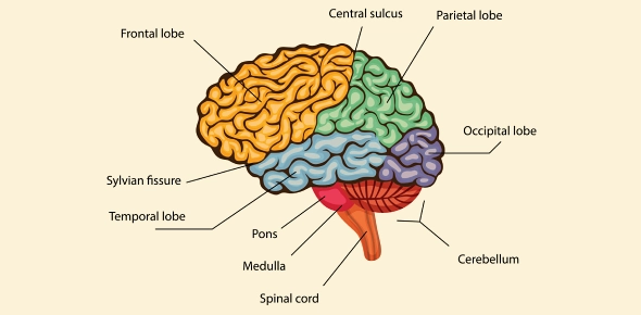
Each cerebral hemisphere is further subdivided into four major lobes: the frontal, parietal, temporal, and occipital lobes. These lobes are regions of the cortex defined by various sulci (grooves) and each is associated with different brain functions.
Let's have a look at these four major lobes:
- Frontal lobe – Controls voluntary movement and executive functions like decision-making, planning, and speech production.
- Parietal lobe – Processes touch, pain, temperature, and spatial awareness.
- Temporal lobe – Handles hearing, language comprehension, memory, and emotion.
- Occipital lobe – Dedicated to visual processing.
Broadly, the frontal lobe focuses on motor control and higher thinking, while the parietal, temporal, and occipital lobes are primarily involved in sensory processing. This classification helps in identifying the functional map of the brain.
The Brainstem (Midbrain, Pons, Medulla)
The brainstem, which connects the brain to the spinal cord, is made up of three parts: the midbrain, pons, and medulla oblongata. It controls many automatic survival functions and acts as a major pathway for information flow.
- Midbrain – Located beneath the cerebrum, it integrates sensory and motor signals. It contains the colliculi, which manage visual and auditory reflexes, and it also helps regulate alertness. Cranial nerves III and IV arise here, mainly controlling eye movements.
- Pons – This middle section links the cerebrum to the cerebellum and medulla. It helps regulate breathing and relays signals between brain regions. The pons houses cranial nerves V–VII, which are responsible for facial sensation, expression, and eye motion.
- Medulla oblongata – The lowest part of the brainstem, it regulates heart rate, blood pressure, and breathing. It is also where many nerve tracts decussate-cross from one side of the brain to the opposite side of the body. The medulla houses cranial nerves VII–XII and governs vital reflexes like swallowing and coughing.
Together with the cerebellum, the pons and medulla form the hindbrain, which anchors the brain to the spinal cord.
(191).jpg)
The Cerebellum
The cerebellum, located beneath the occipital lobes and behind the brainstem, plays a key role in balance, coordination, and fine motor control. It receives input about body position and movement, then adjusts motor signals for smooth execution.
The cerebellum's surface is folded into narrow ridges called folia, which increase its surface area, just like gyri in the cerebrum. Unlike the cerebrum, it works ipsilaterally-each hemisphere controls the same side of the body. This structure ensures coordination, posture, and precision in movement. It doesn't initiate movement but fine-tunes it automatically without conscious effort.
White Matter Pathways: Commissural, Projection, and Association Fibers
The brain contains three major types of white matter fiber tracts, which allow communication between different regions:
- Commissural fibers – Connect the two hemispheres. The corpus callosum is the largest of these, enabling communication between the left and right sides.
- Projection fibers – Link the cerebrum with lower brain regions and the spinal cord. These fibers move information vertically to and from the cortex. A key example is the internal capsule.
- Association fibers – Connect regions within the same hemisphere. Short association fibers link adjacent gyri, while longer ones like the arcuate fasciculus connect different lobes.
These networks allow the brain to coordinate complex tasks and integrate information across regions efficiently.
Gray Matter vs. White Matter (Brain vs. Spinal Cord)
Gray matter consists of neuron cell bodies and dendrites, while white matter is made up of myelinated axons. In the brain, gray matter forms the outer layer (cortex), while white matter lies deeper. In contrast, the spinal cord has white matter on the outside and gray matter on the inside.
This reversed pattern supports their roles: the brain's outer gray matter handles processing and thought, while the inner white matter sends messages. In the spinal cord, outer white matter carries signals up and down, while the inner gray matter controls reflexes and local processing. This organization is crucial for the brain and spinal cord to function as a unified control system.
Rate this lesson:
 Back to top