Radiology: Types, Classification, and Main Medical Imaging Techniques
Lesson Overview
Radiology is a vital branch of medical science that uses imaging techniques to diagnose, monitor, and treat various diseases and conditions within the human body. In this lesson, you will explore the fundamental principles of radiology, including the different types such as X-rays, CT scans, MRI, and ultrasound. You'll also learn about the techniques used to capture and interpret medical images, the role of contrast agents, and safety measures involved in radiologic procedures.
What Is Radiology?
Radiology is a specialized field of medicine dedicated to the study and application of various imaging technologies to diagnose and treat diseases. It involves the use of sophisticated equipment and techniques to produce images of the internal structures of the body, which helps in identifying abnormalities, understanding the extent of an illness, or monitoring the progress of treatment. Radiology plays a critical role in modern healthcare by enabling non-invasive visualization of organs, tissues, and bones, providing vital information that is often unattainable through physical examination alone.
What Are the Types of Radiology?
Radiology is broadly divided into various types based on its purpose, imaging technology, and clinical application. Each type serves a specific role in the diagnosis, treatment, and monitoring of diseases. Below are the main types of radiology:
1. Diagnostic Radiology
Purpose: To visualize the internal structures of the body for disease detection and assessment.
Key Modalities:
- X-ray Radiography: For bones, chest, and joints.
- Computed Tomography (CT): For cross-sectional imaging of organs, trauma, and tumors.
- Magnetic Resonance Imaging (MRI): For detailed images of soft tissues, brain, and spine.
- Ultrasound (Sonography): For real-time imaging of soft tissues and fetal development.
- Nuclear Medicine Scans: For organ function (e.g., thyroid, kidney, bone).
- Mammography: For breast cancer screening.
Applications: Fractures, infections, cancer diagnosis, vascular blockages, neurological disorders.
2. Interventional Radiology (IR)
Purpose: To perform minimally invasive procedures using image guidance.
Techniques Include:
- Angiography: Imaging of blood vessels.
- Biopsies: Image-guided needle sampling of tissue.
- Catheter Insertion: For drainage or drug delivery.
- Embolization: Blocking blood flow to tumors or bleeding sites.
Applications: Cancer treatment, uterine fibroids, aneurysms, abscess drainage.
3. Therapeutic Radiology (Radiation Oncology)
Purpose: To treat disease-especially cancer-using controlled doses of radiation.
Types:
- External Beam Radiation Therapy (EBRT): Uses linear accelerators to target tumors.
- Brachytherapy: Radioactive sources are placed inside or near the tumor.
- Stereotactic Radiosurgery (SRS): High-precision radiation (e.g., for brain tumors).
Applications: Cancer treatment, tumor control, palliative care.
4. Nuclear Radiology
Purpose: Uses radioactive tracers to study organ function and structure.
Imaging Techniques:
- Positron Emission Tomography (PET): Detects metabolic activity.
- Single Photon Emission Computed Tomography (SPECT): Visualizes blood flow and function.
Applications: Cancer staging, heart perfusion, brain disorders, bone diseases.
5. Emergency Radiology
Purpose: Rapid diagnostic imaging in trauma or acute medical situations.
Techniques:
- CT scans for internal bleeding.
- X-rays for fractures.
- Ultrasound for abdominal trauma.
Applications: Accident injuries, stroke, chest pain, abdominal emergencies.
6. Pediatric Radiology
Purpose: Imaging tailored to infants, children, and adolescents.
Features:
- Lower radiation doses.
- Specialized techniques for congenital and developmental conditions.
Applications: Birth defects, bone disorders, infections, pediatric cancers.
7. Breast Imaging
Purpose: Early detection and diagnosis of breast abnormalities.
Techniques:
- Mammography
- Breast ultrasound
- Breast MRI
- Stereotactic biopsy
Applications: Breast cancer screening, evaluation of lumps or discharge.
How Is Radiology Classified?
Radiology is classified based on the type of imaging technique used, the purpose of the imaging, and the method of diagnosis or treatment. This classification helps organize radiologic practices into meaningful categories, making it easier to apply specific tools and procedures depending on clinical needs.
1. Classification by Imaging Technique
| Type | Description | Examples |
|---|---|---|
| Conventional Radiology | Uses X-rays to create 2D images on film or digital detectors. | Chest X-ray, limb fractures |
| Computed Tomography (CT) | Combines X-ray images from different angles to create cross-sectional views. | CT brain scan, abdominal CT |
| Magnetic Resonance Imaging (MRI) | Uses magnetic fields and radio waves to visualize soft tissues. | MRI of brain, spine, joints |
| Ultrasound (Sonography) | Uses high-frequency sound waves for real-time imaging. | Obstetric ultrasound, echocardiography |
| Nuclear Medicine | Involves injecting or ingesting radioactive tracers to examine organ function. | Bone scan, thyroid scan |
| Positron Emission Tomography (PET) | Visualizes metabolic activity using positron-emitting radiotracers. | PET-CT for cancer detection |
| Fluoroscopy | Provides real-time continuous X-ray imaging. | Barium swallow, angiography |
| Mammography | Specialized radiography for breast tissue screening. | Breast cancer detection |
2. Classification by Purpose
| Purpose | Type | Focus |
|---|---|---|
| Diagnostic Radiology | Focused on identifying diseases or injuries through imaging. | X-ray, MRI, CT, PET, ultrasound |
| Interventional Radiology | Uses image guidance to perform minimally invasive procedures. | Biopsies, catheter placements, embolization |
| Therapeutic Radiology (Radiation Oncology) | Uses high-energy radiation to treat diseases, especially cancer. | External beam radiation, brachytherapy |
3. Classification by Body System or Clinical Specialty
| Field | Description |
|---|---|
| Neuroradiology | Imaging of the brain, spine, and nervous system |
| Musculoskeletal Radiology | Bones, joints, and soft tissue imaging |
| Cardiovascular Radiology | Heart and blood vessel imaging |
| Pediatric Radiology | Imaging focused on infants and children |
| Chest Radiology | Lungs, heart, and thoracic cavity |
| Abdominal Radiology | Digestive organs, liver, kidneys, and pelvic structures |
| Breast Imaging | Focused on mammograms and breast ultrasounds/MRI |
| Emergency Radiology | Rapid imaging for trauma or critical care scenarios |
4. Classification by Modality Type
| Modality | Basis of Function |
|---|---|
| Ionizing Radiation | Uses radiation (X-ray, CT, PET, fluoroscopy) |
| Non-Ionizing | Uses magnetic or sound waves (MRI, ultrasound) |
Take This Quiz
What Are the Radiology Procedures and Techniques?
Radiology procedures and techniques are medical imaging methods used to view the inside of the body to diagnose, monitor, or treat various conditions. These techniques vary based on the type of imaging modality, the body part involved, and the clinical objective. Below is a detailed breakdown of the major procedures and techniques used in radiology:
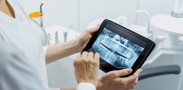
1. X-ray Radiography
- Technique: Uses ionizing radiation to capture static 2D images.
- Procedure: The patient is positioned between the X-ray source and a digital or film detector.
- Common Uses: Bone fractures, chest infections, dental exams.
2. Computed Tomography (CT Scan)
- Technique: Combines multiple X-ray images taken from different angles to produce cross-sectional views.
- Procedure: Patient lies on a motorized table that slides into a rotating gantry; contrast agents may be injected.
- Common Uses: Head trauma, cancer detection, internal bleeding, vascular imaging.
3. Magnetic Resonance Imaging (MRI)
- Technique: Uses strong magnetic fields and radiofrequency pulses to produce detailed images of soft tissues.
- Procedure: Patient lies still in a large tunnel-like scanner; contrast dye (gadolinium) may be used.
- Common Uses: Brain, spine, joints, and organ imaging; neurological and musculoskeletal conditions.
4. Ultrasound Imaging (Sonography)
- Technique: Uses high-frequency sound waves emitted by a transducer to generate real-time images.
- Procedure: A handheld probe is moved over the skin (or inserted into a cavity) with a conductive gel.
- Common Uses: Pregnancy, abdominal organ assessment, heart imaging (echocardiography), thyroid and vascular exams.
5. Fluoroscopy
- Technique: Provides real-time moving X-ray images using continuous radiation.
- Procedure: Often used with contrast media (like barium) to visualize organs in motion.
- Common Uses: GI tract studies, catheter guidance, orthopedic surgery, joint injections.
6. Mammography
- Technique: A specialized low-dose X-ray used to examine breast tissue.
- Procedure: Breast is compressed between two plates to obtain clear images from multiple angles.
- Common Uses: Breast cancer screening, cyst or mass detection.
7. Positron Emission Tomography (PET Scan)
- Technique: Uses radioactive tracers to measure metabolic activity in tissues.
- Procedure: A small amount of radiotracer is injected; the patient then lies in the PET scanner.
- Common Uses: Cancer staging, brain disorders, heart disease.
8. Nuclear Medicine Scans
- Technique: Uses small amounts of radioactive material to evaluate organ function and structure.
- Procedure: Tracer is swallowed or injected; gamma cameras capture its distribution.
- Common Uses: Bone scans, thyroid scans, renal scans, cardiac stress tests.
9. Dual-Energy X-ray Absorptiometry (DEXA Scan)
- Technique: Measures bone mineral density using two X-ray beams of different energy levels.
- Procedure: Patient lies on a table while the scanner passes over the body.
- Common Uses: Osteoporosis diagnosis and monitoring.
10. Interventional Radiology Techniques
- Technique: Minimally invasive procedures performed under imaging guidance (X-ray, CT, ultrasound).
- Procedures Include:
- Angiography – Imaging of blood vessels
- Biopsies – Tissue sampling using needles
- Embolization – Blocking blood flow to tumors
- Catheter Placement – For drainage or medication delivery
What Are the Various Instruments Used in Radiology?
Radiology relies on a range of specialized instruments and machines designed to create detailed images of internal body structures. Each instrument serves a unique purpose based on the type of imaging required and the area being examined. Below is a classification of the major instruments used in radiology:
1. X-ray Machine
- Function: Produces X-rays to create 2D images of bones and dense tissues.
- Components: X-ray tube, detector/film, control panel.
- Common Uses: Fracture detection, chest imaging, dental exams.
2. Computed Tomography (CT) Scanner
- Function: Uses X-rays and computer processing to generate cross-sectional images (slices).
- Components: Gantry (rotating ring), X-ray tube, detectors, computer system.
- Common Uses: Head injuries, internal bleeding, tumor identification.
3. Magnetic Resonance Imaging (MRI) Scanner
- Function: Uses strong magnetic fields and radio waves to produce high-resolution images of soft tissues.
- Components: Magnet, gradient coils, RF coils, patient table.
- Common Uses: Brain scans, spinal cord imaging, joint assessments.
4. Ultrasound Machine
- Function: Uses high-frequency sound waves to create real-time images of internal organs.
- Components: Transducer (probe), control panel, display screen.
- Common Uses: Pregnancy monitoring, abdominal organ examination, heart (echocardiography).
5. Fluoroscopy Unit
- Function: Provides continuous X-ray imaging (real-time) to observe motion or guide procedures.
- Components: X-ray source, fluorescent screen or digital detector, image intensifier.
- Common Uses: GI tract studies, catheter placements, joint injections.
6. Mammography Unit
- Function: Specialized X-ray machine for imaging breast tissue.
- Components: X-ray tube, compression paddle, image receptor.
- Common Uses: Breast cancer screening and diagnostics.
7. Positron Emission Tomography (PET) Scanner
- Function: Detects gamma rays emitted by radiotracers to evaluate metabolic and biochemical activity.
- Components: Radiotracer injector, gamma camera, computer system.
- Common Uses: Cancer staging, brain function analysis, heart disease detection.
8. Dual-Energy X-ray Absorptiometry (DEXA)
- Function: Measures bone mineral density using low-dose X-rays.
- Components: X-ray generator, detectors, patient table.
- Common Uses: Osteoporosis diagnosis.
9. C-Arm Machine
- Function: Mobile fluoroscopy unit used during surgical procedures.
- Components: C-shaped arm with X-ray source and detector.
- Common Uses: Orthopedic surgeries, vascular procedures.
10. Radiographic Film and Digital Detectors
Take This Quiz
- Function: Capture and store images produced by radiation exposure.
- Types: Traditional film, computed radiography (CR), digital radiography (DR).
- Common Uses: All X-ray-based modalities.
Take This Quiz
(97).jpg)
Conclusion
This radiology lesson has provided an in-depth exploration of the history, classifications, procedures, and key figures that have shaped radiology into what it is today. We have also learned about the essential role of anatomy in interpreting radiological images, the importance of radiation safety protocols, and the innovative use of advanced radiology software in clinical practice. Furthermore, we have highlighted the diverse instruments used in various imaging techniques and how radiologic technology is applied to guide minimally invasive procedures, monitor treatment efficacy, and support preventive medicine.
Rate this lesson:
 Back to top