Skeletal Muscle: Structure, Functions, and How Contraction Works
Lesson Overview
- What Is Skeletal Muscle and Why Is It Critical to Human Function?
- How Is Skeletal Muscle Structured Microscopically?
- What Are the Key Proteins Involved in Skeletal Muscle Contraction?
- How Does Skeletal Muscle Contraction Occur Mechanistically?
- What Is the Functional Difference Between Origin and Insertion?
- What Are Tendons and How Do They Transmit Muscle Force?
- How Are Antagonistic and Synergistic Muscles Organized?
- What Are the Histological Differences Between Muscle Types?
- What Are Myofibrils and Their Role in Muscle Function?
- How Are Muscle Actions Coordinated Through Innervation?
- What Is Muscle Fatigue and How Does It Affect Performance?
- What Conditions or Diseases Affect Skeletal Muscle Function?
When athletes suffer performance drops or patients struggle to regain mobility, the issue often lies in the skeletal muscle. This lesson explains how muscle fibers, contractions, and attachments all work together. Students will explore muscle structure, function, and coordination-building a strong foundation in anatomy and real-world physiology.
What Is Skeletal Muscle and Why Is It Critical to Human Function?
Skeletal muscle is a form of striated muscle tissue under voluntary control, primarily responsible for body movement, posture, and heat production. This muscle type connects to bones through tendons and contracts upon stimulation by motor neurons.
- Skeletal muscles constitute approximately 40% of the body mass in adults.
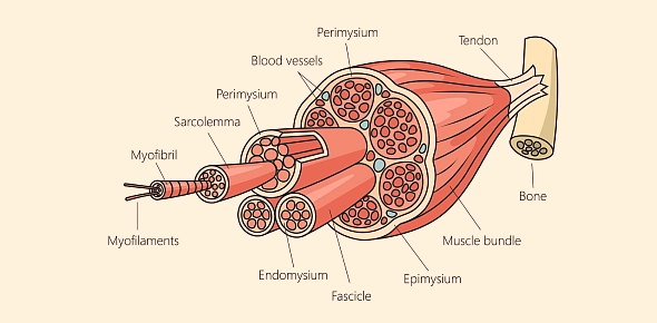
- Each muscle is made up of bundles of muscle fibers (cells) enclosed in connective tissue.
- These fibers are multinucleated and contain specialized organelles called myofibrils, responsible for contraction.
(127).jpg)
How Is Skeletal Muscle Structured Microscopically?
Skeletal muscle exhibits a highly organized internal structure, allowing precise and strong contractions.
- Muscle fibers contain cylindrical structures called myofibrils.
- Myofibrils are composed of sarcomeres, the functional units of contraction.
- Each sarcomere includes alternating light (I bands) and dark (A bands) due to actin and myosin filament alignment.
| Structure | Function |
| Sarcolemma | Muscle cell membrane; conducts action potentials |
| Sarcoplasmic reticulum | Stores and releases calcium ions for contraction |
| Myofibrils | Contractile units composed of sarcomeres |
What Are the Key Proteins Involved in Skeletal Muscle Contraction?
Skeletal muscle contraction depends on the interaction between thick and thin filaments:
- Myosin is a thick filament that binds ATP and interacts with actin.
- Actin is a thin filament anchored at the Z-line, providing binding sites for myosin.
- Troponin and tropomyosin regulate access to binding sites on actin.
Upon calcium binding, troponin causes tropomyosin to shift, exposing the actin-myosin binding sites, initiating contraction.
How Does Skeletal Muscle Contraction Occur Mechanistically?
The contraction process follows the sliding filament theory:
- An action potential reaches the neuromuscular junction.
- Acetylcholine is released, depolarizing the sarcolemma.
- Calcium is released from the sarcoplasmic reticulum.
- Calcium binds to troponin, shifting tropomyosin.
- Myosin binds actin, performs a power stroke, and releases ADP.
- ATP binds to myosin, causing detachment and re-cocking.
This cycle continues as long as calcium and ATP are available.
What Is the Functional Difference Between Origin and Insertion?
Each skeletal muscle has two points of attachment:
- Origin is the stationary attachment during contraction.
- Insertion is the movable attachment pulled toward the origin.
For example, in biceps brachii:
- Origin: Scapula.
- Insertion: Radius.
This anchoring allows force transmission through tendons, which are fibrous tissues connecting muscle to bone.
What Are Tendons and How Do They Transmit Muscle Force?
Tendons are dense connective tissues composed primarily of collagen fibers. These fibers have tensile strength and transmit mechanical force from contracting muscles to bones.
- Tendons are non-elastic but capable of withstanding high tension.
- The tendon-bone interface is called the enthesis.
- Injuries like tendinitis or ruptures can impair muscle function and mobility.
Understanding tendon mechanics is essential for rehabilitation and orthopedic health.
How Are Antagonistic and Synergistic Muscles Organized?
Muscles typically work in groups to execute movement:
- Agonist: Primary muscle performing a movement.
- Antagonist: Muscle that opposes the agonist's action.
- Synergist: Assists the agonist by reducing unnecessary motion.
Example:
- Biceps brachii (flexion) vs. Triceps brachii (extension) as antagonistic pair.
- Brachialis acts as a synergist to biceps during elbow flexion.
Proper coordination among these groups ensures efficient and controlled motion.
What Are the Histological Differences Between Muscle Types?
Three types of muscle tissue exist: skeletal, cardiac, and smooth.
| Feature | Skeletal Muscle | Cardiac Muscle | Smooth Muscle |
| Control | Voluntary | Involuntary | Involuntary |
| Striations | Present | Present | Absent |
| Nuclei | Multinucleated | Single nucleus | Single nucleus |
| Location | Attached to bones | Heart walls | Hollow organs, vessels |
| Unique Feature | Fast, forceful contraction | Intercalated discs | Slow, sustained contraction |
Smooth muscle lacks regular sarcomere alignment, explaining its non-striated appearance【158†source】.
What Are Myofibrils and Their Role in Muscle Function?
Myofibrils are cylindrical organelles made of repeating sarcomeres containing actin and myosin. They occupy most of the intracellular space of muscle fibers and are essential for contraction.
- Each myofibril is surrounded by sarcoplasmic reticulum.
- They are aligned in register, giving rise to the striated appearance.
- Disruption or degeneration of myofibrils results in muscle weakness.
Sarcomere length and arrangement determine force output and contractile velocity.
How Are Muscle Actions Coordinated Through Innervation?
Motor neurons control muscle contraction via neuromuscular junctions:
- Each muscle fiber receives input from a motor neuron at a motor endplate.
- Acetylcholine release triggers an action potential in the sarcolemma.
- The action potential travels via T-tubules to the sarcoplasmic reticulum.
- Calcium release initiates the contractile cycle.
Motor units (one neuron and the fibers it innervates) vary in size:
- Small motor units control fine movements (e.g., eyes).
- Large motor units control gross movements (e.g., thighs).
What Is Muscle Fatigue and How Does It Affect Performance?
Muscle fatigue results from the decline in muscle's ability to generate force. Causes include:
- Accumulation of lactic acid.
- Depletion of ATP and glycogen.
- Failure of neural input.
Fatigue can be:
- Central (nervous system origin).
- Peripheral (muscle cell origin).
Proper hydration, nutrition, and training help delay fatigue and improve recovery.
What Conditions or Diseases Affect Skeletal Muscle Function?
Several disorders impact skeletal muscle health:
- Muscular dystrophy: Genetic condition causing progressive muscle wasting.
- Myasthenia gravis: Autoimmune disorder impairing neuromuscular transmission.
- Rhabdomyolysis: Breakdown of muscle tissue releasing myoglobin into the bloodstream.
- Tendinitis: Inflammation of tendon, impairing muscle-to-bone transmission.
Early diagnosis and targeted therapies can improve quality of life in affected individuals.
Rate this lesson:
 Back to top