|
General senses |
|
large group of touch receptors |
| |
|
special senses |
|
taste, smell, sight, hearing, and balance |
| |
|
Localized receptors |
|
confined to head region; receptors aren't free endings of sensory neurons |
| |
|
-Specialized receptor cells |
|
neuron-like epithelial cells or small peripehral neurons |
| |
|
Taste |
|
-gustation
-receptors classifed as "chemoreceptros" that respond to food dissolced in saliva fluids |
| |
|
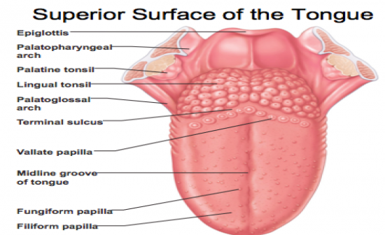
Superior surface of tongue |
|
-stratified squamons epithelium
-filiform papillae
-fungiform papillae
-vallate papillae
-sulcus terminalis: marks border between mouth and pharynx
-Lingual tonsil: covers posterior 1/3rd of tonge that lies in oropharynx |
| |
|
Superior surface of tongue image |
|
|
| |
|
Filiform papillae |
|
-most numerous papillae on tongue are small and concial pointed in shape and line up in parallel rows which enable tonge to grasp and manipuate food; DON'T contain tate buds |
| |
|
Fungiform papillae |
|
mushroom shapd and spread over anterior 2/3rd of tongue on surface; contain taste buds
|
| |
|
Vallate papillae |
|
V-shaped row bordering the posterior third of the tongue and directly anterior to the terminal sulcus (sulcus terminalis) groove; contain taste buds |
| |
|
Taste buds |
|
-contain taste receptors
-collection of 50-100 epithelial cells
-contain two major cell types
-> Gustatory epithelial cells
-> Basal epithelial cells
-contain long microvilli (gustatory hairs): extend trhough a taste pore to the surface of the epithelim
-Cells in tastebuds replaced every 7-10 days |
| |
|
Taste Sensation and Gustatory Pathway |
|
-Five basic qualities of taste: sweet, sour, salty, bitter, and umami; "umami" elicited by glutamate
-Taste map is a myth
-all taste modalities can be elicited from all areas of tongue containg taste buds (fungiform and vallate papillae) |
| |
|
Gustatory Pathway |
|
-Taste information reaches the cerebral cortex primarily through the facial (CN VII), glossopharyngeal (CN IX) and vagus nerve (CN X)
-bitter taste receptors ahve been found in stomach
-Gustatory sensory neurons synapse in which solitary nuclues of medulla from which impulses are transmitted to teh thalamus and ultimately to the gustatory area of the cerebral cortex in insula |
| |
|
Smell |
|
-olfaction
-Receptors: classified as "chemoreceptors" that resond to airborne chemical that dissolve in fluids of nasal mucosa |
| |
|
Olfaction |
|
-olfactory receptors are part of the olfactory epithelium
-Olfactory epithelium is pseudostratified simple columan and contains three main cell types
->olfactory sensory neurons
->supporting epithelial cells
->basal epithelia cells |
| |
|
Smell: cell body to epithelium |
|
-cell bodies of olfaactory sensory neurons are locted in olfactory epithelium
-have an apical dendrite that projects to the epithelial surface and ends in a know from which olfactory cilia radiate
-Olfactory cilia act as receptive structures for smell
-mucus captures and dissolves odor molecules |
| |
|
Smell: cell body to brain |
|
-Gather into bundles of axon filaments of the olfactory nerve
-pas through the cribiform plate of the ethmoid bond
-attach to olfactory bulbs and synapse with mitral cells (output cells)
-Mitral cells transmit impulses along the olfactory tract to:
1. Limbic system
2. Piriform lobe of cerebral cortex |
| |
|
Anosmia |
|
-absence of smell due to injury, colds, allergies, or zinc deficiency |
| |
|
Uncinate fits |
|
-distortion of smells or olfactory hallucinations
-> often result of irritation of olfactory pathways
-after brain surgery or head trauma |
| |
|
Eye & vision |
|
-70% of all sensory receptros in body are in eyes
-40% of cerebral cortex is involved in processing visual infromation
-Anterion 1/6th of eye's surface is visible while rest is positioned in boney eye sockey |
| |
|
Accessory structures of eye |
|
-eyebrows: coarse hairs on superciliary arches
-eyelids (palpebrae): separated by palpebral fissure
-> meet at medial and lateral angles (canthi)
-> lacrimal caruncle: reddish elevation at the medial canthus
->tarsal plates: connective tissue within the eyelids
->tarsal glands: modified sebaceious glands
-lacrimal puntum
-lacrimal canaliculus
-nasolacrimal duct
-inferior meatus |
| |
|
conjunctiva |
|
transparent mucous membrane
-palperbral (tarsal) conjunctiva
-Bulbar conjunctiva
-conjunctival sac |
| |
|
lacrimal apparatus |
|
keeps surface of eye moist |
| |
|
lacrimal gland |
|
produces lacrimal fluid |
| |
|
lacrimal sac |
|
fluid empties into nasal cavity |
| |
|
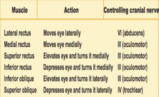
Extrinsic Eye muscles |
|
-Six extrinsic eye muslces that control movement of eye
->originate in walls of orbit
->insert on outer surface of eyeball
->annular ring: origin of four rectus muscles
-6 muscles are:
-> lateral rectus and medial rectus
->superior rectus and inferior recuts
->superior oblique and inferior oblique |
| |
|
Summary of eye muscle actions & innnervating CNs |
|
|
| |
|
Anatomy of eyeball |
|
-components of eye
-> protect and support photoreceptors
->Gather, focus, and process light into precise images
-Anterior pole: most anterior part of eye
-Posterior pole: most posterior part of eye
-External walls: composed of three tunics
-Internal cavity: contains fluids (humors) |
| |
|
Anterior and posterior segments of eyes |
|
-lens and ciliary zonules divide eye into anterior and posterior segements |
| |
|
Medial view of eye |
|

|
| |
|
Posterior segement of eye |
|
-Fillled with vitreous humor
-clear, jelly-like substance
-transmits light
-supports the posterior surface of lens
-helps maintain intraocular pressure |
| |
|
Anteriro segement of eye contains both anterior and posterior chambers |
|
-Anterior chamber: between cornea and iris
-Posterior chamber: between iris and lens
*both chambers are filled with aqueous humor |
| |
|
Aqueous humor |
|
-formed from a filtrate of the capillaries in the ciliary processes
-flows from posterior chamber through pupil into the anterior chamber
-reabsorbed into venouse blood by the sclereal veous sinus
-renewed continuously
-provides nutrients to the lens oand cornea |
| |
|
glaucoma |
|
-increaed intraocular pressure in anterior segment
-occurs when aqueous humor drains more slowly than it forms
-causes compression on retina and optic nerve which can lead to blindness |
| |
|
Lens |
|
-thichk, transparent, biconvex disc
->held i place by ciliary zonule
-Lens epithelium: covers anterior surface of lens
-Elongated lens fibers form bulk of lens
->new lens fibers are continuously added
-> lens enlarges throughout life |
| |
|
Eye as an optical device |
|
-structures in eye bend light rays
-light rays converge on retina at a single focal point
-light bending structures (refractory media) are the lens, cornea, humors |
| |
|
Accomodation |
|
curvature of lens is adjustable which allows fro focusing on nearby objects |
| |
|
3 layers of wall of eye |
|
-Fibrous layer: slcera & cornea
-Vascular leyer: choroid, ciliary body, & iris
-Sensory layer: pigmented layer, neural layer |
| |
|
Fibrous layer |
|
-mot external layer of eyeball and is composed of dense connective tissue in 2 differnt regions: sclera and cornea |
| |
|
sclera |
|
-posterior 5/6ths of tunic
->white, opaque region
->provides shape and anchor for eye muscles |
| |
|
cornea |
|
-anterior 1/6th of fibrous tunic
-epithelium (outer layer)
-thick layer of dense collagen-rich connective tissue
-endothelium (inner layer)
-cornea is avascular, but gets oxygen from air in front and nutrients from aqueous humor behind
-is richly suppled with sensory nerve endings, most of which are pain receptors |
| |
|
Limbus |
|
junction between sclera and cornea
|
| |
|
scleral venous sinus |
|
allows aqueous humor to drain |
| |
|
Vascular Layer is middle coat of eyeball and is composed of |
|
-choroid
-ciliary body
-iris |
| |
|
Choroid |
|
-vascular
-darly pigmented membrane
-forms posterior 5.6ths of vascual tunic
-brown color: from melanocytes
-prevents scattering of light rays within the eye
-choroid corresponds to teh arachnoid and pia maters |
| |
|
Ciliary body |
|
-thickened ring of tissue, which encircles lens
-ciliary body consists chiefly of smooth muscle called ciliary muscle which acts to focus the lens
->ciliary processes: posterior surface of ciliary body
->ciliary zonule (suspensory ligament) is a halo of fine fibrils attached around entire circumference of the lens |
| |
|
Iris |
|
-visible colored part of eye
-attached to ciliary body and positioned anterior to the ciliary body
-composed of smooth muscle |
| |
|
Pupil |
|
-round, central opening
->sphincter pupilae muscle (circularly arranged)
-dilator pupillae muscle (radially arranged)
*act to vary size of pupil
-pupillary light reflex
-> protective response of pupil constriction when a bright light is flashed in the eye |
| |
|
Inner layer of eye |
|
-Retina: think outer pigmented layer and a thick inner neural layer
-optic nerve |
| |
|
Regional specialiaitons of Retina |
|
-Neural layer ends oat teh posterior margin of the ciliary body and this junction is called "ora serrata retinae"
-Macula lutea: containsm mostly cones
-Fovea centralis: contains only cones and i region of highest visual acuity
-Optic disc: blind spot |
| |
|
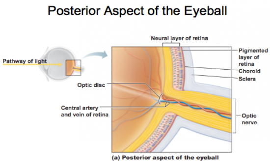
Posterior Aspect of Eyeball |
|
|
| |
|
Inner Layer (Retina) |
|
-deepest tunic; composed of 2 layers
-> pigmented layer: single outer layer of melanocytes
-> neural layer: most inner layer with a sheet of nervous tissue
-contains 3 main types of neurons
->photoreceptor cells (rods and cones)
-bipolar cells
-ganglion cells
-4th cell type is interneurons (amacrine and horizontal cells) which process visual information |
| |
|
Retina neural layer of Inner Layer |
|
-Photoreceptor cells: signal bipolar cells
-Bipolar cells signal ganglion cells ot generate nerve impulses
-axons from ganglion cells run along internal surface of retina
->converge posteriorly to form optic nerve |
| |
|
Rod Cells |
|
photoreceptro that is more sensitive to light and allow vision in dim light |
| |
|
cone cells |
|
operates best in bright light and enable high-acuity, color vision |
| |
|
Photoreceptors |
|
-Rods and cones have an inner and outer segment
-outer segments are receptors regions that contain light absorbing pigments
-light particles modify the visual pigment and generate a nerve impulse |
| |
|
Visual Pathway to the cerebral cortex |
|
-pathway begins at the retina
-> light activates photoreceptors
-> photoreceptors signal bipolar cells
-> bipolar cells signal ganglion cells
-> axons of ganglion cells exit eye as optic nerve
-axons of ganglion cells exit the eye at the optic nerve
-axons extending from medial (nasal) side of retina run ipsilaterlally into optic tract
-Optic tract send axons to the lateral geniculate nucleus of thalamus and synapse with thalamic neurons
-Fibers of optic radiation reach the primary visual cortex |
| |
|
Visual Pathways to other parts of the brain |
|
-some axons from optic tracts (branch to midbrain)
-> superior colliculi: controls extrinsic eye muscles
-> pretectal nuclei: mediate pupillary eye refelexes
-Other branches form optic tracts
-Branch to the suprachiasmatic nucleus of hypothalamus which processes visual input to help synchronize daily biorythyms witht he daylight-darkness cycle |
| |
|
Blood supply of retina |
|
-Retina receives blood from two sources
-> outer third of the retina is supplied by capillaires in the choroid
-inner 2/3 of retina is serviced by central artery and vein of retina |
| |
|
Cataracts |
|
-Lens becomes opaque which results in blindness; typically develops in people over 50 |
| |
|
Age-related macular degeneration (AMD) |
|
involves the buildup of deposits in retina which causes loss of vision in macula which si the center of the visual field, typically develops in people over 50 years of age
|
| |
|
trachoma |
|
contagioius infectrion from chlamydia trachomatis of the conjunctiva which causes the eyelids to become distorted and inverted which results in eyelashes scraping against cornea which can lead to blindess from corneal scarring |
| |
|
Hearing & equilibrium |
|
-Ear: receptor organ for hearing and equilibrium
-composed of 3 main regions
->outer ear: functions in hearing
-> middle ear: functions in hearing
-Inner ear: function in hearing and equilibrium |
| |
|
Outer (external) ear |
|
Aurice (pinna): helps direct sounds
-external acoustic meatus (external ear canal): lined with skin, hairs, sebaceious glands, and ceruminous glands
-Tympanic membrane: forms the boundary between the external and middle ear |
| |
|
Ear infections |
|
-Otitis externa: infection and inflammation of skin linin external acoustic meatus (externa ear canal); "swimmer ear" can be a cause of this problem
-Otitis media" infection of tympanic membrane |
| |
|
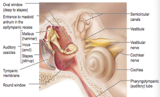
Middle ear |
|
composed of
-Tympanic cavity (forms lateral wall)
->small, air-filled space
->located within petrous portion of temporal bone
-Medial wall is penetrated by
-> oval window
-> round window
-Pharyngotympanic tube (auditory or eustachian tube) linkes middle ear and pharynx |
| |
|
Ear ossicles |
|
smallest bones in the body
-> malleus: attaches to the ear drum
->incus; between malleus & stapes
-> stapes: vibrates against the val window
-Tensor tympani and stapedius muscles
-> two tiny skeletal muscles in the middle ear |
| |
|
Internal ear |
|
-internal ear: also called labyrinth
-lies within the petrous portion of the temporal bone
-membranous labyrinth is filled with a clear fluid -endolymph
-bony labyrinth is filled with perilymph which is continuos with CSF fluid |
| |
|
Bony labyrinth in internal ear |
|
-bony labyrinth in internal ear is a cavity consiting of three parts
->semicircular canals
->vestiblue
->cochlea |
| |
|
Middle & internal ear image |
|
|
| |
|
Membranous Labyrinth in Internal ear |
|
-series of membrane-walled sacs and ducts
-fit within the bony labyrinth
-consists of three main parts
->semicircular ducts within semicircular canals
->utricle and saccule within vestibule
->cochlear duct within cochlea |
| |
|
Inner ear structures |
|
-Semicircular canals (bony labyrinth) contain semicircular ducts (membranous labyrinth) which facilitates equilibium related to rotational acceleration of the head
-Vestiblue (bony labyrinth) contains utrricle and saccule (membranous labyrinth) which facilitates equilibrium related to static equiilibrium and linea acceleration of the head
-Cochlea (bony labyrinth) contains cochelar duct (membranous labyrinth whcih facilitates hearing |
| |
|
Cochlea |
|
-spiral chamber in bony labyrinth
-from its attachmetn on the vestibule it coils around a pillar of bone: the modiolus
-Osseous spiral lamina: spiral of bone in modiolus
-Cochlear nerve runs through the core of the modiolus
-cochlear duct lies betwen two chambers: scala vestibuli & the scala tympani
-scala vestibuli is connected with the oval window
-Helicotrema is region at apex of cochlea where scala vestibuli and the scala tympani are continuous |
| |
|
vestibular membrane |
|
roof of cochlear duct |
| |
|
basilar membrane |
|
floor of cochlear duct
|
| |
|
spiral organ (of corti) |
|
receptor epithelium for hearing which is supported by basilar membrane |
| |
|
cochlea: spiral organ of corti |
|
-cochelar duct (scala media): contains receptors for hearing
-spiral organ of corti: receptor epithelium for hearing which consits of
-> supporting cells are columan epithelium
-inner and outer hair cells (receptor cells)
-> one row of inner hair cells are the receptors that transmit vibraitons of the basilar membrane
-> three rows of outer hair cells actively tune the cochlea and amplify the signal
-at the apex of hair cells, the tips of the hairs (stereocilia) are embedded in the gel-like tectorial membrane
-at their base, the hair cell synapse with the sensory fibers of the cochlear nerve |
| |
|
Auditory pathway from organ of corti in cochlea |
|
-spiral organ of corti in cochlea
-spiral gagnlion of cohclear nerve
-cochlear nuclei (CN VIII) of medulla
-superior olivary nucleus of medula-pons junction
-medial geniculate nucleus of thalamus
-primary auditroy cortex of temporal lobe |
| |
|
Deafness |
|
-Conduction deafness
-> sound vibrations cannot be conducted to the inner ear
--> ruptred tympanic membrane, otitis meida, otosclerosis
-Sensorineural deafness
-> results from damage to any part of the auditory pathway |
| |
|
Vestibule |
|
-Central part of the bony labyrinth
-lies medial to the middle ear
-Utricle and saccule are two egg-shaped parts of the membranous labyrinth
-Utricle and saccule both contain a "macula" spot of sensory epithelium
-Utricles is continuous with the semicircular ducts
-Saccule is continuous with coclear duct |
| |
|
Vestibule: macula |
|
-contains receptor cells
-monitor the position of head when the head is still (static equilibrium) and when head is moving straight ahead (linear acceleration)
-contains columna supporting cells
-receptor cell: called "hair cells"
base of hair cells synapse with vestibular nerve
-apex of hair cells contains many sterocilia and one kinocilium
-tips of these hair cells are embedded in otolithic membrane which is a jelly-like disc that contains crystals of calcium carbonate called "otoliths" |
| |
|
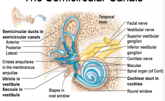
Semicircular canals |
|
-lie posterior and lateral to the vestibule
-anterior and posterior semicircular canals
-> lie in the vertical plane at right angles
-Lateral semicircular canal
->lies in the horizontal plane |
| |
|
Image of semicircular canals |
|
|
| |
|
Semicircular duct |
|
snakes through each semicircular canal |
| |
|
Membranous ampulla |
|
(swelling in semicircular duct)
-located within bony ampulla houses a structure called "crista ampullaris"
->cristae contain receptor cells of rotaitonal acceleration
->epithelium contains supporting cells and receptor hair cells (kinocilium and stereocilium)
-hairs of these hair cells project into a tail, jelly-like mass called a cupula, that resembleas a pointed hat |
| |
|
Equilibrium Pathway |
|
-transmits infomration on the position and movement fo the head
-information on equilibrium is relatyed from receptors in the bestibule an semicircualr canals of the inner ear throught the vestibular nuclei in the medulla to the flocculonodular lobe in the cerebellu |
| |
|
Motion sickness |
|
-carsickness, seasickness
-> popular theory for a cause, a mismatch of sensory inputs
|
| |
|
Meniere's Syndrome |
|
-equilibrium greatly distrubed
-> excessive amounts of endolymph in the membranous labyrinth |
| |
