|
Cyst Definition |
|
- an abnormal cavity, lined by epithelium, containing fluid or semi-solid material
- can be developmental and get larger in time
can be found in soft tissue as well as bone |
| |
|
Cyst Classification |
|
- based on the source of the epithelium (odontogenic cyst, non-odontogenic cyst)
- based on their location (soft tissue, intrabony) |
| |
|
Odontogenic Cyst |
|

- a cyst which the lining of the lumen is derived from epithelium produced during tooth development
- can come from different parts of the odontogenic structures (Rests of Malassez, reduced enamel epithelium, dental lamina, etc)
- Also Classified on their location |
| |
|
Periapical Cyst Definition |
|

- odontogenic cyst from the Rests of Malassez, this is one of the most common cysts
- located at the root apex of the tooth
- the tooth must be infected or inflammed which will extend to the apical area
- an odontogenic cyst of inflammatory origin that is preceeded by a chronic PA granuloma and stimulation of rests of Malassez |
| |
|
Periapical Cyst Characteristics |
|

- PA cyst is always a radiolucency, but we cannot make a diagnosis based on the radiograph alone
- we cannot determine a cyst from a chronic inflammation (apical granuloma) without histological evaluation
- Failed RCT will lead to a PA cyst
- one requirement is having a necrotic/infected tooth (if the tooth is intact, this will be some other pathology) |
| |
|
Periapical Cyst Treatment |
|

- apical surgery is necessary to remove the cyst and then retrofill the lesion
- if the tooth is not salvagable, then we must remove the retained root and remove the cyst otherwise it will turn into a residual cyst
- note that histologically there are inflammatory cells and a capsule surrounding an epithelial layer with a lumen on the interior |
| |
|
Rushton Bodies |
|

- these are eiosinophilic bodie which result from degenerative changes in cells in some (not all) of the cystic lesions
- this is an indication of the longevity of a chronic lesion |
| |
|
Residual Cyst |
|
- this develops from an untreated radicular cyst
- we must curate the lesion otherwise it will lead to a residual cyst |
| |
|
Dentigerous Cyst (Follicular Cyst) |
|

- this is an odontogenic cys (from the developmental structures of the tooth) which surrounds the crown of an impacted tooth
- caused by fluid accumulation between the reduced enamel epithelium and enamel surface resulting in a cyst
- this is a pericoronal cyst of an impacted tooth
- lined by non-keratinized epithelium (could be inflammed or not) |
| |
|
Eruption Cyst |
|

- this is an odontogenic cyst with the histologic features of a dentigerous cyst that surrounds a tooth's crown that has erupted through the bone but NOT the soft tissue
- clinically is visible as a soft fluctuant mass on the alveolar ridges
- found in children who are in the process of erupting their teeth
- possible to have a blue-ish color due to trauma and bleeding with pigments (hemosiderin) |
| |
|
Odontogenic Keratocyst (OKC) |
|
- cyst derived from the remnants, rests of the dental lamina with a biologic behavior similar to a benign neoplasm
- distinct lining of 6-10 cells in thickness that exhibits a basal cell layer of palisaded cells and a surface of corrugated parakeratin
- they can be found anywhere in the mouth, when around the root of a tooth they can cause resorption of the bone |
| |
|
Odontogenic Keratocyst Characteristics |
|

- this has histologically keratinized epithelium, thus if you look at the lumen of the OKC it will be filled with a cheezy material (keratin)
- have a uniform thickness with a flat basal membrane, no rete pegs, and a 7-8 cell layer
- not usually inflamed, not irritated
- also might see a micro-cyst or a daughter/satellite cyst in the cell wall of the OKC (leading to an increased probability of recurrence |
| |
|
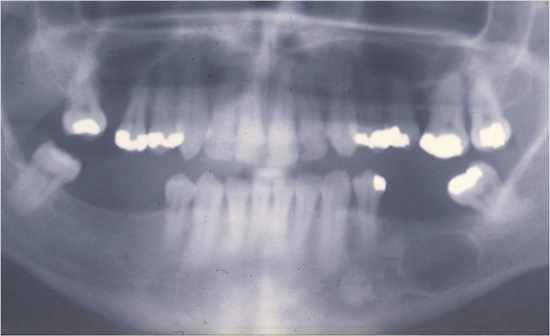
OKC in Basal Cell Carcinoma Syndrome |
|

- most of the OKC are single and thus if there are multiple present we must think of BCC syndrome
- also present with bifid ribs, pitting of the skin, CV problems, intracranial tumors
- complexity of the syndrome leads to a treatment of individual problems but not of the syndrome
- dentist is usually first to diagnose syndrome due to the presence of multiple OKC |
| |
|
Lateral Periodontal Cyst |
|

- a slow-growing, non-expansive developmental odontogenic cyst derived from one or more rests of the dental lamina containing an embryonic lining of 1-3 cuboidal cells and distinctive focal thickenings (plaques)
- found between two adjacent teeth in the bone, common in the posterior mandible, rarely change shape and lined by NON-keratinized |
| |
|
Butryoid Odontogenic Cyst |
|

- a type of lateral periodontal cyst that shows a multilocular growth pattern (looks grapelike)
- these are many cystic cavities which are lined by the same epithelium as the lateral periodontal cysts |
| |
|
Gingival Cyst of the Adult |
|

- a small developmental odontogenic cyst of the gingival soft tissue derived from the rests of the dental lamina
- contains a lining of the embryonic epithelium of cuboidal cells and distinctive focal thickenings similar to the lateral periodontal cysts
- this lesion is in the soft tissue rather than in bone
- asymptomatic, slow growing, but may gradually cause bone resorption due to pressure |
| |
|
Dental Lamina (Gingival) Cyst of the Newborn |
|
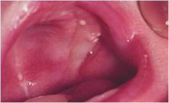
- also called Epstein Pearl or Bohn's Nodule
- uncommon superficial raised nodules on the edentulous ridge of infants that resolve without treatment
- derived from the rests of the dental lamina and consisting of keratin-producing epithelial cells |
| |
|
Glandular Odontogenic Cyst (Sialo-Odontogenic Cyst) |
|

- an unusually large solitary or multi-locular odontogenic cyst most likely from the rests of dental lamina
- stratified squamous epithelium containing numerous mucus-secreting cells
- these present as radiolucencies, several mucus-producing cavities right next to each other |
| |
|
Paradental Cyst |
|

- a cyst of uncertain origin found primarily on the distal or facial aspect of a vital mandibular third molar consisting of intensely inflamed CT and epithelial lining |
| |
|
Nasopalatine Duct Cyst |
|

- an intraosseous developmental cyst of the midline of the anterior palate, derived from the islands of epithelium remaining after the closure of the embryonic nasopalatine duct
- lesion (when large) tend to be symptomatic due to pressure and inflammation
- these are around vital teeth which respond normally to testing
- cysts may have nerves and vessels due to size |
| |
|
Nasolabial Cyst |
|

- a developmental cyst of the soft tissue of the anterior mucobuccal fold beneath the ala of the nose
- most likely derived from the remnants of the inferior portion of the nasolacrimal duct
- requires surgical removal, non-odontogenic cyst seen in the upper lip area |
| |
|
Lymphoepithelial Cyst |
|
- a cyst with a lumen lined by a keratinizing stratified squamous epithelium and a capsule containing multiple normal lymphoid follicles and a dense accumulation of the normal lymphocytes
- smaller lesions in the oral cavity, larger aspect in the lateral neck |
| |
|
Oral Lymphoepithelial Cyst |
|

- a lymphoepithelial cyst commonly located intraorally on the posterior lateral tongue and the anterior floor of the mouth
- the cavity is lined by keratininzed epithelium with lymphoid tissue under the surface (germinal center) |
| |
|
Cervical Lymphoepithelial Cyst |
|

- also called a branchial cleft cyst
- an unusually large lymphoepithelial cyst located on the lateral aspect of the neck
- upon removal we see a cavity with lymphoid tissue with the same tissue as that in the lymph nodes, this should be removed |
| |
|
Thyroglossal Duct Cyst |
|

- a cyst located above the thyroid gland and beneath the base of the tongue
- lumen is lined with a mixture of epithelial cell types derived from the remnants of the embryonic thyroglossal tract, often containing thyroid tissue in the capsule
- upon removal it resembles thyroid tissue histologically |
| |
|
Dermoid Cyst |
|

- cyst of the midline of the upper neck or anterior floor of the mouth of young patients, derived from the remnants of the embryonic skin
- consists of a lumen lined by a keratinized stratified squamous epithelium and containing one or more skin appendages (hair, sweat, sebasceous glands)
- lesions that are lined by epithelium containing another misplaced (ectopic) demoid substance
- location of the cyst depends on its relation to the mylohyoid muscle |
| |
|
Epidermoid Cyst |
|

- a cyst of skin with a lumen lined by keratinizeing stratified squamous epithelium, usually filled with keratin and without skin appendages in the capsule wall
- similar to the dermoid cyst, but we do not see appendages
- keratin filled cyst with no appendages |
| |
|
Surgical Ciliated Cyst of the Maxilla |
|

- an intrabony cyst located near the floor of the maxillary sinus lined by a pseudostratified ciliated columnar epithelium, caused by implantation of normal mucus secreting sinus epithelium during surgery
- in maxilla when surgical perforation happens placing a portion of the epithelium into the bone of the maxilla resulting in a cyst |
| |
|
Heterotopic Oral Gastrointestinal Cyst |
|
- a rare and unusual developmental cyst commonly found in the tongue or floor of the mouth of infants or young children
- histologically looks like it has features of the GI |
| |
