


Related Flashcards
Related Topics


Cards In This Set
| Front | Back |
|
1) What are the three types of muscle?
2) The mechanism of __________ is similar between all three types. |
1) skeletal (striated)cardiacsmooth
2) The mechanism of muscle contraction is similar between all three types. |
|
The muscles of the skeletal system are part of which function of the nervous system???
|
Motor Output!!!!
|
|
1)describe some characteristics of a muscle cell??come on you can do it!!!
2) why are skeletal muscles striated? |
1) multinucleated, striated, they undergo DNA replication but DO NOT divide, these nuclei make the proteins that make up myofibrils (i.e. myosin and actin)
2) the arrangement of the myosin (thick filaments, ATPase) and actin (thin filaments) cause the appearance of the striations because of how they are arranged in each sarcomere |
|
Describe the arrangement of the sarcomere!! (this is very important to know because it is crucial to contraction)
|
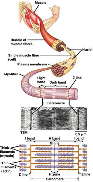 - composed of myosin and actin filaments!!- The Z-line anchors thin filaments- H zone is the middle, where there are no myosin heads- A band is where there are actin filaments and myosin- I band is where there are only actin filaments; between Z-line this image from slide 4 should clarify the structural appearance for you!! |
|
How do the structures of the muscle come together? (i.e what are the building blocks of the muscular tissue)
|
 Myosin and actin make up sarcomeres |sarcomere makes my myofibril |Myofibrils make up a muscle fiber (cell) |Bundles of muscle fibers make up the muscular tissue (the bicep in the pic from slide 4) |
|
How does muscle contraction work in relation to the myosin-actin interactions involved??
|
 Look at slide 6 |
|
How does muscle contraction impact shape of sarcomere and what occurs generally?
|
Myosin head hyrolyze ATP as an energy source for moving the thin filaments (actin) inwardthe Z-line shifts inward to the center of the sarcomere and the as each of the 100,000 plus sarcomere of a muscle cell strinks, you get a contraction!!!
|
|
The sliding filament hypothesis!! Explain it. (i know these may seem redundant but hey I am literally adding all of my notes as he said it) The generalized version of contraction and the sliding hypothesis are one in the same.
|
During contraction, both the H bands and I bands decrease in
length as the myosin thick filament
“pulls” the actin thin filament which is anchored by the Z-line toward
the M-line. Each sarcomere shortens by
about 0.5 um.
|
|
When the ATP in your muscles are used, what disease are you most likely going to develop??
|
Rigor mortis
|
|
How does Calcium regulate Muscle Contraction??
|
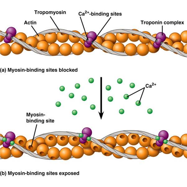 The myosin binding site on actin is blocked by tropomyosin in the relaxed muscle. When calcium binds to troponin the tropomyosin shifts, exposing the binding site on actin. Note: Calcium binds to troponin when calcium levels are high and this is regulated by the nervous system, because the nervous system regulate calcium levels in muscle fibrils |
|
Can muscle cells generate action potentials?
what type of ion channels are on the plasma membrane on muscle fibers? Is each muscle in contact with a motor neuron? Y/N |
Yes
voltage-gated channels Yes!! |
|
How does the nervous system regulates contractions in the muscle in relation to how calcium is released into muscle?
|
Calcium is stored in the muscle’s endoplasmic reticulum
(called sarcoplasmic reticulum).
Release of calcium is regulated by depolarization of the muscle cell caused by release of acetylcholine from the motor neuron. The depolarization is carried into the interior of the muscle by the Transverse Tubules (T-tubules), which are associated with the sarcoplasmic reticulum. When the depolarization (action potential) hits the Sarcoplasmic reticulum, calcium is released and it binds to troponin |
|
The overview of how the muscle and nervous system work together to bring about a contraction!!!
|
Look at you all done with muscles and shit!!***Pat yourself on the back because you only have three more lectures left!!****
|
