|
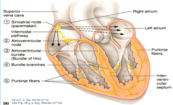
What are the specialised cells that initiate the electrical impulse in the heart. |
|
Sinoatrial node (pacemarker) is a group of specialised muscle cells under the superior vena cava in the top of the right atria |
| |
|
How is the electrical pathway initiated? |
|
The sinoatrial node opens its sodium channels the fastest of any cardiac muscle cell which starts an action potential |
| |
|
What is the second node of the electrical pathway and what does it do? |
|
There is an internodal pathway and the second node is the Atrioventricular node, which slows down the pulse allowing time for the ventricles to fill before contraction. |
| |
|
What is the third part of the electrical pathway of the heart? |
|
Bundle of His, Atrioventicular bundle. |
| |
|
What is the fourth part of the electrical pathway of the heart? |
|
Bundle branches which branch off from the bundle of His, down the Septum to the apex |
| |
|
What is the last part of the electrical pathway of the heart? |
|
The Purkinje fibres conduct the impluse from the apex toward the artia of the heart. This contracts the heart in an upward direction squeezing blood through the semilunar valves into the arteries with force (pressure).
These fibres spread the impulse to every other cell they are attached to. |
| |
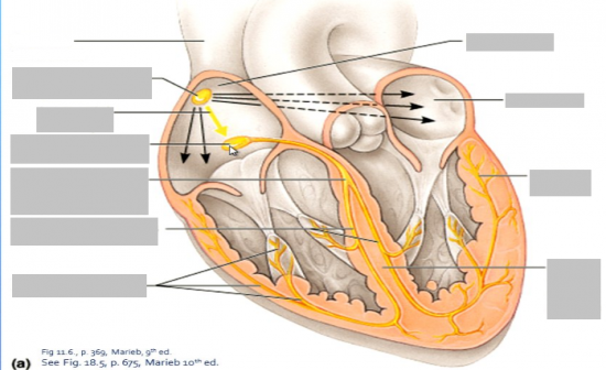
Label this diagram |
|
|
| |
|
Explain the initation and conduction of electrical impulses in the heart (SHORT ANSWER) |
|
Intrinsic Cardiac Conduction
*The hear can generate its own action potentials by opening its own sodium channels
* Auto-rhythmic cardiac cells generate action potentials which set the heart's nasic rhythm
* The cells of the sinoatrial (SA) node do this more rapidly than any other cardiac muscle cells,
* this is why it is called the pacemaker. Even though other cells are capable, but just not as quickly.
* The autonomic nervous system coordinates and modifies the pace (extrinsic cardiac conduction)
In order to contract, and pump blood, impulses spread through specialised muscle cells from the
*Sino-atrial (SA) node
* Atrioventricular (AV) node
* AV bundle (bundle of His)
* Right and left bundle branches
* Purkinje fibres
Sino-atrial node depolarises leads to an impulse
The impulse spreads across both atria to AV node
Atrial cells then contract
Impulse slowed through the AV node which allowed the blood to reach the ventricles before the impulse
Impluse tracels across the AV bundle, down the left and right bundle branhes to Purkinje fibres
Ventricles depolarise and then contract
Contraction begins in the apex and spreads upwards. |
| |
|
What is a polarised cell? |
|
negative in and positive out
Potassium in and Sodium out |
| |
|
What is a depolarised cell? |
|
positive in and negative out
Sodium in and Potassium in |
| |
|
What is repolarised? |
|
negative in and positive out
Sodium in and Potassium out |
| |
|
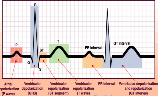
Draw and label a graph of an ECG |
|

|
| |
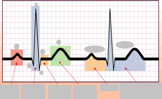
Label this diagram |
|
|
| |
|
How does an ECG work? |
|
Electrocardiogram
In order to contract, cardiac muscle cells move Sodium ions and then Potassium ions aross their plasma membrane (the same as for skeletal muscle contraction)
This leads to an electrical signal that can be recorded (on the ECG)
Producing the PQRST complex waves
ECG recorded the hearts electrical activity and monitors cardiac changes
It cannot show contractions
The P wave = atrial depolarization
The ORS complex = ventricular depolarization (artial repolarisation is hidden within this complex)
The T wave = ventricular repolarisation |
| |
|
What is happening at each wave of an ECG? |
|
Electrocardiogram
P wave = atrial depolarisation
QRS cpmlex = ventricular depolarisation (artial repolarisation is hidden in the complex)
T wave = ventricular repolarisation |
| |
|
What is the cardiac cycle? |
|
All events associated with the pressure and volume changes within the heart during one heart beat
Cardiac cycle:
Systole = contraction
Diastole = relaxation
Cardiac cycle = All events associated with pressure and volume changes within the heart during one heart beat
|
| |
|
What are the events during the cardiac cycle? |
|
Mid-late - diastole (ventricular filling - relaxation of the muscles then artia contraction or systole)
Ventricle systole (ventricle contraction) - pushing the blood out of the ventricles
Early diastole (ventricular relaxation then ventricular filling) |
| |
|
Describe mid-to-late diastole. |
|
(when the heart is relaxed)
Blood flows passively through the open AV valves into the ventricles
80% of vlood enters the ventricles this way
The SL valves are closed
Artia depolarise which leads to contractions which leads to the remaining 20% of blood being pumped into the ventricles via atria systole
The impluse now spreads via the AV node, through the AV bundle and bundle branchs to the Pukinje fibres |
| |
|
Describe ventricular systole |
|
As the bentricles fill the AV valves drift upwards
The ventricles then depolarise and begin to contract
The pressure increases as the SL valves are closed
The AV valves snape close which leads to the lub
heart sound.
At same time the artia repolarise which leads to atrial diasotle and passive filling
increasing ventricular press, less than the pressure in the great vessels, which leads to the opening of the semilunar valves
Blood is then pumped into the aorta and pulmonary arteries |
| |
|
Describe early diastole |
|
Ventricular repolarisation which leads to a decrease in ventricular pressure
The blood in the great vessels then falls back towards the ventricles, snapping the semilunar valves shut which is the dub sound.
During ventricular systole the atria have been in distole, filling with blood and the intra-artial pressure as been increased |
| |
