|
Define Exchange Surface |
|
An exchange surface is a specialised area that is adapted to make it easier for molecules to cross from one side of the surface to the other. |
| |
|
What substances do living cells need? (6) |
|
- oxygen for aerobic respiration
- glucose as a source of energy
- proteins for growth and repair
- fats to make membranes and to be a store of energy
- water
- minerals to maintain their water potential and to help enzyme action and other aspects of metabolism |
| |
|
What features do good exchange surfaces have in common? |
|
- large surface area to provide more space for molecules to pass through
- thin barrier to reduce diffusion distance
- fresh supply of molecules on one side to keep the concentration high
- removal of required molecules on the other side to keep the concentration low
|
| |
|
List 4 exchange surfaces in living organisms |
|
1) small intestine (where nutrients are absorbed)
2) liver (where levels of sugars in the blood are adjusted)
3) root hairs of plants (where water and minerals are absorbed)
4) hyphae of fungi (where nutrients are absorbed) |
| |
|
Define Gaseous exchange |
|
Gaseous exchange is the movement of gases by diffusion between an organism and its environment across a barrier such as the alveolus wall. |
| |
|
How does gaseous exchange occur in the lungs? |
|
Gases pass both ways through the thin walls of the alveoli. Oxygen passes from the air in the alveoli to the blood in the capillaries. Carbon dioxide passes from the blood to the air in the alveoli. |
| |
|
Why do the lungs have a large surface area? |
|
The large surface area provides more space for molecules to pass through. |
| |
|
Why do the lungs have a barrier permeable to oxygen and carbon dioxide? |
|
The plasma membranes that surround the thin cytoplasm of the cells form the barrier to exchange. These readily allow the diffusion of oxygen and carbon dioxide. |
| |
|
Why do the lungs have a thin barrier to reduce diffusion distance? |
|
- the alveolus wall is one cell thick
- the capillary wall is one cell thick
- both walls consist of squamous cells
- the capillaries are in close contact with the alveolus walls
- the capillaries are so narrow that the red blood cells are squeezed against the capillary wall, making them closer to the air in the alveoli and reducing the rate at which they flow past in the blood
- the total barrier to diffusion is only two flattened cells thick |
| |
|
Why must the lungs produce surfacant? |
|
To reduce the cohesive forces between the water molecules. Without the surfacant, the alveolus would collapse due to the cohesive forces between the water molecules lining the air sac. |
| |
|
Why is a steep diffusion gradient needed in the lungs? |
|
For diffusion to be rapid. |
| |
|
What is meant by a steep diffusion gradient? |
|
This means having a high concentration of molecules on the supply side of the exchange surface and a low concentration on the demand side. |
| |
|
How is a steep duffusion gradient maintained? |
|
A fresh supply of molecules on one side is needed to keep the concentation there high, and a way of removing the molecules from the other side is needed to keep the concentration there low. |
| |
|
How is a diffusion gradient maintained in the lungs? |
|
This is achieved by the action of the blood transport system and the ventilation movements.
The blood brings carbon dioxide from the tissues to the lungs. This ensures that the concentration of carbon dioxide in the blood is higher than that in the air of the alveoli. It also carries oxygen away from the lungs, This ensures that the concentration of oxygen in the blood is kept lower than the concentration in the air inside the alveoli. The heart pumps the blood along the pulmonary artery to the lungs. In the lungs, the artery divides up to form finer and finer vessels. These eventually carry blood into tiny capillaries that are only just wide enough for a red blood cell to squeeze through. These capillaries lie over the surface of the alveoli.
The breathing movements fo the lungs ventilate the lungs. They replace the air used with fresh air. This brings more oxygen into the lungs and ensures that the concentration of oxygen in the air of the alveolus remains higher than the concentration in the blood. Ventilation also removes air containing carbon dioxide from the alveoli. This ensures that the concentration of carbon dioxide in the alveoli remains lower than that in the blood. |
| |
|
Outline inspiration (inhaling) |
|
- Diaphragm contacts to become flatter and pushes digestive organs down
- External intercostal muscles contract to raise ribs
- Volume of chest cavity increases
- Pressure in chest cavity drops below atmospheric pressure
- Air moves into lungs |
| |
|
Outline expiration (exhaling) |
|
- Diaphragm relaxes and is pushed up by displaced organs underneath
- External intercostal muscles relax and ribs fall
- Volume of chest cavity decreases
- Pressure in lungs increases and rises above atmospheric pressure
- Air moves out of lungs |
| |
|
What 5 requirements must the airways meet? |
|
1) larger airways must be large enough to allow sufficient air to flow without obstruciton
2) they must divide into smaller airways to deliver air to all the alveoli
3) the airways must be strong enough to prevent them collapsing when the air pressure inside is low
4) they must be flexible, to allow movement
5) they must be able to stretch and recoil |
| |
|
What is the difference between the trachea and bronchi? |
|
The bronchi are narrower |
| |
|
List the 4 features of the trachea and bronchi |
|
1) Much of the wall consists of cartilage
2) The cartilage is in the form of incomplete rings in the trachea, but is less regular in the bronchi
3) On the inside surface of the cartilage is a layer of glandular tissue, connective tissue, elastic fibres, smooth muscle and blood vessels.
4) The inner lining is an epithelium layer that has two types of cell. Most of the cells have cilia. This is called cilliated epithelium. Among the ciliated cells are goblet cells. |
| |
|
Describe the bronchioles |
|
They are much narrower than the bronchi.
Larger bronchioles may have some cartilage.
The wall is made mostly of smooth muscle and elastic fibres.
Very small bronchioles have clusters of alveoli at their ends. |
| |
|
What is the role of cartilage? |
|
Cartilage plays a structural role. It supports the trachea and bronchi, holding them open. |
| |
|
What is the role of smooth muscle? |
|
The smooth muscle can contract. When it contracts it constricts the airway and makes the lumen of the airway narrower. Constricting the lumen can restrict the flow of air to and from the alveoli. |
| |
|
What is the role of elastic fibres? |
|
When smooth muscle contacts, it reduces the diameter of the lumen of the airway. The smooth muscle cannot reverse this effect. When the airway constricts, it deforms the elastic fibres in the loose tissue. As the smooth muscle relaxes, the elastic fibres recoil to their original size and shape. This helps to dilate the airway. |
| |
|
What is the role of goblet cells and glandular tissue? |
|
Goblet cells and glandular tissue secrete mucus. Mucus traps tiny particles from the air. These particles may include pollen and bacteria. Removing the bacteria will reduce the risk of infection. |
| |
|
What is the role of ciliated epithelium? |
|
This consists of ciliated cells. The cilia on these cells (hair like projections) move in a synchronised pattern to waft mucus up the airway to the back of the throat. The mucus can then be swallowed and the acidity in the stomach will kill any bacteria. |
| |
|
What is tidal volume? |
|
Tidal volume is the volume of air moved in and out of the lungs with each breath when you are at rest. Normally 0.5dm3 |
| |
|
What is vital capacity? |
|
Vital capacity i sthe largest volume of air that can be moved into and out of the lungs in any one breath. Normal 5dm3 |
| |
|
What is residual volume? |
|
Residual volume is the volume of air that always remains in the lungs, even after the biggest possible exhalation. Normally 1.5dm3 |
| |
|
What is dead space? |
|
Is the air in the bronchioles, bronchi and trachea. |
| |
|
What is inspiratory reserve volume? |
|
Inspiratory reserve volume is how much more air can be breathed in over and above the normal tidal volume when you take in a big breath. You call on this reserve when exercising. |
| |
|
What is expiratory reserve volume? |
|
Expiratory reserve volume is how much more air can be breathed out over and above the amount that is breathed in a tidal volume breath. |
| |
|
How does a spirometer work? |
|
A spirometer consists of a chamber filled with oxygen that floats on a tank of water. A person breathes in from a disposable mouthpiece attached to a tube connected to the chamber of oxygen. Breathing in takes oxygen from the chamber, which then sinks down. Breathing out pushes air into the chamber, which then floats up.
The movements of the chamber are recorded using a dataloger so that a spirometer trace can be produced. |
| |
|
Why is soda lime used in measuring oxygen uptake? |
|
Soda lime is used to absorb the carbon dioxide that is exhaled. This causes the volume of gas in the spirometer to go down. |
| |
|
Look at spirometer trace |
|
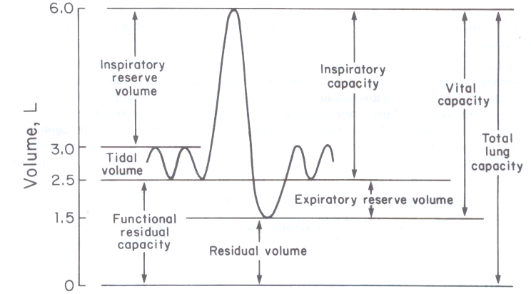
 |
| |
|
Define transport |
|
Transport is the movement of oxygen, nutrients, hormones, waste and heat around the body. |
| |
|
What three factors affect the need for a transport system? |
|
- size
- surface area to volume ratio
- level of activity |
| |
|
What 5 things will and effecient/effective transport system include? |
|
- a fluid or medium to carry nutrients and oxygen around the body (blood)
- a pump to create pressure that will push the fluid around the body (heart)
- exchange surfaces that enable nutrients and oxygen to enter the blood and to leave it again where they are needed
- tubes or vessels to carry the blood
- two circuits (one to pick up oxygen and another to deliver oxygen to the tissues) |
| |
|
What sort of circulatory system to fish have? |
|
Single circulatory system |
| |
|
How does a single circulatory system work in fish? |
|
The blood flows from the heart to the gills and then on to the body before returning to the heart. |
| |
|
What sort of circulatory system do mammals have? |
|
A double circulatory system |
| |
|
How does the double circulatory system work? |
|
One circuit carries blood to the lungs to pick up oxygen. This is the pulmonary circulation. The other circuit carries the oxygen and nutrients around the body to the tissues. This is the systemic circulation. The mammalian heart is adapted to form two pumps - one for each circulation. Blood flows through the heart twice for each circulation of the body. |
| |
|
What happens in the single circulatory system of fish? |
|
- the blood pressure is reduced as blood passes through the tiny capillaries of the gills
- this means it will not flow very quickly to the rest of the body
- this limits the rate at which oxygen and nutrients are delivered to respiring tissues.
Fish are less active than mammals and do not maintain their body temperature, so they need less energy. Their single circulatory system delivers oxygen and nutrients quickly enough for their needs, so for them it is efficient. |
| |
|
What happens in the double circulatory system of mammals? |
|
- the heart can increase the pressure of the blood after it has passed through the lungs, so blood flows more quickly to the body tissues
- the systemic circulation can carry blood at a higher pressure than the pulmonary circulation
- the blood pressure must not be too high in the pulmonary circulation, otherwise it may damage the delicate capillaires in the lungs.
Mammals are active animals and maintain their body temperature. Both the energy for activity and the heat needed to keep the body warm require enough energy from food. The energy is released from food in the process of respiration. To release a lot of energy, the cells need good supplies of both nutrients and oxygen. |
| |
|
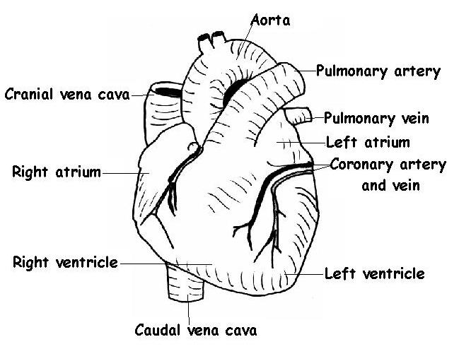
Define the heart |
|
The heart is a muscular pump that creates pressure to propel blood through the arteries and around the body. |
| |
|
What does the right side of the heart do? |
|
Pumps deoxygenated blood to the lungs to be oxygenated. |
| |
|
What does the left side of the heart do? |
|
Pumps oxygenated blood to the rest of the body. |
| |
|
What forces blood along the arteries? |
|
The heart squeezes the blood, putting it under pressure. The pressure forces the blood along the arteries. |
| |
|
Look at diagram of heart |
|
|
| |
|
What do the coronary arteries do? |
|
They carry oxygenated blood to the heart muscle itself. |
| |
|
What do the atria do? |
|
These recoeve blood from the major veins. |
| |
|
Where does deoxygenated blood from the body flow? |
|
From the vena cava into the atrium. |
| |
|
Where does oxygenated blood from the lungs flow? |
|
From the pulmonary vein into the left atrium. |
| |
|
Where does blood flow from the atria? |
|
Blood flows down through the atrioventricular valves into the ventricles. |
| |
|
What happens when the ventricles contract? |
|
The valves fill with blood and remain closed. This ensures that the blood flows upwards into the major arteries and not back into the atria. |
| |
|
What is the septum and what does it do? |
|
A wall of muscle that seperates the ventricles from each other. This ensures that the oxygenated blood in the left side of the heart and the deoxygenated blood in the right side are kept seperate. |
| |
|
What do the semilunar valves do? |
|
They are at the base of the major arteries and prevent blood returning to the heart as the ventricles relax. |
| |
|
Why is the muscle of the atria very thin? |
|
Because these chambers do not need to create much pressure. Their function is to push the blood into the ventricles. |
| |
|
Why are the walls of the right venticle thicker than the walls of the atria? |
|
This enables the right ventricle to pump blood out of the heart. |
| |
|
Why are the walls of the right ventricle thinner than those of the left? |
|
The right ventricle pumps deoxygenated blood to the lungs. The lungs are in the chest cavity beside the heart, so the blood does not need to travel very far. Also, the lungs contain a lot of very fine capillaries that are in close contact with the walls of the alveoli. The alveoli walls are very thin and there is very little or no tissue fluid. So the capillaries are not supported and could easily burst. The pressure of the blood must be kept down to prevent the capillaries in the lungs bursting. |
| |
|
Why are the walls of the left venrticle much thicker than the right? |
|
The blood from the left ventricle is pumped out through the aorta and needs sufficient pressure to overcome the resistance of the systemic circulation. |
| |
|
Define cardiac cycle |
|
The cardiac cycle is the sequence of events in one heartbeat. |
| |
|
Describe the filling phase (1) |
|
While both the atria and the ventricles are relaxing, the internal volume increases and blood flows into the heart from the major veins. The blood flows into the atria, then through the open atrioventricular valves and into the ventricles. This phase of the cycle is called diastole. |
| |
|
Describe atrial contraction (2) |
|
The heart beat starts when the atria contract. Both right and left atria contract together. The small increase in pressure created by this contraction helps to push blood into the ventricles. This stretches the walls of the ventricles and ensures they are full of blood. Contraction of the atria is called atrial systole. Once the ventricles are full the begin to contract. Blood fills the atrioventricular valve flaps causing them to snap shut. This prevents blood returning to the atria. |
| |
|
Describe ventricular contraction (3) |
|
Now there is a short period when all four heart valves are closed. The walls of the ventricles contract. This is called ventricular systole. This raises the pressure in the ventricles very quickly. The contraction starts at the apex (base) of the heart so this pushes the blood upwards towards the arteries. The semilunar valves open and blood is pushed out of the heart. The contraction only lasts for a short time. Then the ventricle walls relax allowing the heart to start filling again. |
| |
|
How do the atrioventricular valves work? |
|
When the venticle walls relax and recoil after contracting, the pressure in the ventricles drops below the pressure in the atria. This causes the atrioventricular valves to open. Blood entering the heart flows straight through the atria and into the venticles. The pressure in the atria and the ventricles slowly rises as they fill with blood. The valves remain open while the atria contract. As the ventricles begin to contract, the pressure of the blood in the ventricles rises. When the pressure rises above that in the atria, the blood starts to move upwards. This movement fills the valve pockets and keeps them closed. This prevents the blood flowing back into the atria. |
| |
|
How do the semilunar valves work? |
|
When the ventricles start to contract, the pressure in the major arteries is higher than the pressure in the ventricles. This means that the semilunar valves are closed. As the ventricles contract, the pressure inside rises very quickly because the blood cannot escape. Once the pressure in the ventricles rises above the pressure in the aorta and pulmonary arteries, the semilunar valves are pushed open. The blood is under very high pressure, so is forced out of the ventricles in a poweful spurt. Once the ventricle walls have finished contracting, the heart muscle strarts to relax. Elastic tissue in the walls of the ventricles recoils to stretch the muscle out again and return the ventriclet to its original size. This causes the pressure in the ventricle to drop quickly. As it drops below the pressure in the major arteries, the semilunar valves are pushed closed by the blood starting to flow back towards the ventricles and collecting in the pockets of the valves. This prevents blood returning to the ventricles. |
| |
|
What is the sound of the heart? |
|
The sound of the heart is made by the valves closing. - the first sound (lub) is made by the atrioventricular valves closing as the ventricles start to contract. - the second sound (dup) is made by the semilunar valves closing as the ventricles start to relax. |
| |
|
Define sionatrial node |
|
The SAN is the heart's pacemaker. It is a small patch of tissue that sends out waves of electrical excitation at regular intervals to initiate contactions. |
| |
|
Define Purkyne tissue |
|
Purkyne tissue is specially adapted muscle fibres that conduct the wave of excitation from the AVN down the septum to the ventricles. |
| |
|
Why is heart muscle described as myogenic? |
|
Because it can initiate its own contraction. |
| |
|
How does the heartbeat start? (1) |
|
At the top of the right atrium, near the point where the vena cava empties blood into the atrium, is the SAN. There is a small patch of tissue that generates electrical activity. The SAN initiates a wave of excitation at regular intervals. |
| |
|
Desctibe how contaction of the atria works (2) |
|
The wave of excitation quickly spreads over the walls of both atria. It travels along the membranes of the muscle tissue. As the wave of excitation passes, it causes the cardiac muscle cells to contract. This is atrial systole.
At the base of the atria is a disc of tissue that cannot conduct the wave of excitiation. So the excitation cannot spread directly to the ventricle walls. At the top of the inner-ventricular septum is another node - the atrio-ventricular node (AVN). This is the only route through the disc of non-conducting tissue. The wave of excitation is delayed in the node. This allows time for the atria to finish contracting and for the blood to flow down into the ventricles before they begin to contract. |
| |
|
Descirbe how contraction of the ventricles works (3) |
|
After this delay, the wave of excitation is carried away from the AVN and down specialised conducting tissue. This is Purkyne tissue and it runs down the inter-ventricular septum. At the base of the septum, the wave of excitation spreads out over the walls of the ventricles. As the excitation spreads upwards from the base (apex) of the ventricles, it causes the muscles to contract. This means that the ventricles contract from the base upwards, pushing blood up to the major arteries at the top of the heart. |
| |
|
What are electrocardiograms and how do they work? |
|
ECG's are used to measure the activity heart. Sensors are attached to the skin. Some of the electrical activity generated by the heart spreads through the tissues next to the heart and onwards to the skin. The sensors on the skin pick up the electrical excitation created by the heart and convert this into a trace. |
| |

What is this?
 |
|
A normal ECG
- P shows excitation of the atria
- QRS indicates excitation of the ventricles
- T shows diastole |
| |
|
Define open circulatory system. |
|
In an open circulatory system the blood is not always in vessels. |
| |
|
Define closed circulatory system. |
|
In a closed circulatory system the blood always remains within vessels. |
| |
|
Why don't all animals have an open system? |
|
Open systems work for insects as they are small and they don't rely on the blood to transport oxygen and carbon dioxide.
Larger organisms rely on blood to transport oxygen and carbon dioxide. In an open system blood remains at low pressure and flows slowly. This would not be sufficient to meet the needs of the muscles in a large, active animal. |
| |
|
What happens in closed circulatory systems? |
|
The blood stays entirely inside vessels. Tissue fluid bathes cells and tissues. This means blood can be pumped at a higher pressure and flow quickly. It means oxgyen and nutrients can be delivered quickly and wastes can be removed quickly. Fish have a closed single circulatory system. |
| |
|
Describe arteries. |
|
Arteries carry blood away from the heart. The blood is at high pressure so the wll must be able to withstand that pressure.
- The lumen is relatively small to withstand that pressue.
- The wall is relatively thick and contains collagen to give it strength to withstand pressure
- The wall has elastic tissue that allows the wall to stretch and then recoil when the heart pumps.
- The wall contains smooth muscle that can contract and constrict the artery. The constriction narrows the lumen.
- The endothelium is folded and can unfold when the artery stretches. |
| |
|
Describe veins. |
|
Veins carry blood back to the heart. The blood is at low pressure and the walls do not need to be thick.
- The lumen is relatively large to ease the flow of blood.
- The walls have thinner layers of collagen, smooth muscle and elastic tissue. They do not need to stretch and recoil, and are not actively constricted to reduce blood flow.
- The main feature of veins is that they contain valves to help the blood flow back to the heart and prevent it flowing in the opposite direction. As the walls are thin, the vein can be flattened by the action of the surrounding skeletal muscle. Pressue is applied to the blood, forcing the blood to move along in a direction dictated by the valves. |
| |
|
Describe capillaries. |
|
Capillaries have very thin walls. They allow exchange of materials between the blood and cells of tissues via the tissue fluid.
- The walls consist of a single layer of endothelial cells that reduced the diffusion distance for the materials being exchanged.
- The lumen is very narrow. This ensures that red blood cells are squeezed as they pass along the caplllaries. This helps them give up their oxygen because it presses them close to the capillary wall, reducing the diffusion path to the tissues. |
| |
|
Define blood |
|
Blood is held in the heart and blood vessels. |
| |
|
Define tissue fluid |
|
Tissue fluid bathes the cells of induvidual tissues. |
| |
|
Define lymph |
|
Lymph is help within the lymphatic system. |
| |
|
Describe blood. |
|
Blood consists of blood cells in a watery liquid called plasma. The plasma contains many dissolved substances. The cells include red and white blood cells as well as platelets. |
| |
|
Describe tissue fluid and its role |
|
Tissue fluid is similar to blood but does not contain plasma proteins or as many cells. The role of tissue fluid is to transport oxygen and nutrients from the blood to the cells, and to carry carbon dioxide and other wastes to the blood. |
| |
|
Explain how tissue fluid is formed |
|
The fluid that leaves the capillary is known as tissue fluid. This fluid surrounds the body cells, so exchange of gases and nutirents can occur across the cell surface membranes. This exchange occurs by diffusion and facilitated diffusion. Oxygen and nutrients enters the cells; carbon dioxide and other wastes leave the cells. |
| |
|
Explain how the fluid returns to the blood. |
|
The tissue fluid itself has some hydrostatic pressure and this pushes the fluid back into the capillaries. Both the blood and tissue fluid contain solutes which give them a negative water potential. The water potential of the fluid is less negative than that of the blood. This means that water moves back into the blood from the tissue fluid via osmosis, down the water potential gradient. At the venuous end of the capillary the blood has lost its hydrostatic pressure. The combined effect of hydrostatic pressure and the osmotic force moves the fluid into the capillary. It carries with it any waste substances. |
| |
|
Describe the formation of lymph. |
|
Lymph is similar to tissue fluid and contians the same solutes. |
| |
|
What is haemoglobin? |
|
Haemoglobin is a complex protein with 4 subunits. Each subunit consists of a polypeptide chain and a haem group. The heam group has a high affinity for oxygen. |
| |
|
How does haemoglobin take up oxygen? |
|
Oxygen molecules diffuse into the blood plasma and enter the red blood cells. Here they are taken up by haemoglobin. This takes the oxygen molecules out of solution and so maintains a steep diffusion gradient. This diffusion gradient allows more oxygen to enter cells. |
| |
|
How is oxygen released by heamoglobin? |
|
Cells need oxygen for aerobic respiration. Oxyhaemoglobin must be able to release the oxygen. This is called dissociation. |
| |
|
Describe and explain haemoglobin and oxygen transport |
|
The ability of haemoglobin to take up and release oxygen depends on the amount of oxygen in the surrounding tissues. This is called partial pressure (oxygen tension).
Haemoglobin can take up oxygen so it creates and S shaped curve called the oxyhaemoglobin dissociation curve. |
| |
|
What is fetal heamoglobin? |
|
Has a higher affinity for oxygen than adult haemoglobin. It must be able to absorb oxygen from the fluid in the mother's blood. This reduces oxygen tension within the blood fluid which makes the maternal haemoglobin release oxygen. Therefore the dissociation curve for fetal haemoglobin is to the left of the curve for adult haemoglobin. |
| |
|
How is carbon dioxide transported? |
|
5% dissolved directly into plasma
10% combined with haemoglobin to form carbaminohaemoglobin
85% is transported in the form of hydrogencarbonate ions. |
| |
|
How are hydrogencarbonate ions formed? |
|
CO2 enters the red blood cell and is combined with water to form carbonic acid. The enxyme carbonic anhydrase ensures that this occurs. The carbonic acid dissociates to hydrogen ions and hydrogencarbonate ions. |
| |
|
Describe the Bohr effect |
|
The Bohr effect results in oxygen being more readily released where more carbon dioxide is produced from respiration. |
| |
|
Define xylem |
|
Xylem transports water up the plant |
| |
|
Define phloem |
|
Phloem transports sugars and other assimilates up and down the plant |
| |
|
Why do plants need transport systems? |
|
So that all of the cells can recieve enough water and nutrients in order to survive. |
| |
|
What substances do plants need to move? |
|
- water and soluble minerals (these travel upwards in xylem tissue) - sugars (these travel up and down in phloem tissue) |
| |
|
What are the vascular tissues? |
|
xylem and phloem |
| |
|
What is the vascular bundle? |
|
The xylem and phloem are found together in vascular bundles. These bundles often contain other types of tissue that give the bundle some strenght and help to support the plant. |
| |
|
Describe xylem and phloem in the stem. |
|
The vascular bundles are found near the outer edge of the stem. The xylem is found towards the inside of each vascular bundle. The phloem is found towards the outside of the bundle. In between the xylem and phloem is a layer of cambium. The cambium is a layer of meristematic cells that divide to produce new xylem and phloem. |
| |
|
Describe xylem and phloem in the leaf |
|
The vascular bundles form the midrib and veins of a leaf. A dicotyledon leaf has a branching network of veins that get smaller as they spread away from the midrib. Within each vein, the xylem can be seen on top of the phloem. |
| |
|
Describe the structure of xylem |
|
Xylem tissue consitsts of tubes to carry the water and dissolved minerals, fibres, to help support the plant and living parenchyma cells. |
| |
|
What are xylem vessels? |
|
These are long cells with thick walls that have been inpregnated by lignin. - The lignin waterproofs the walls of the cells. As a result the cells die, this leaves a long column of dead cells with no contents. - the lignin strenghtens the vessel and prevents it from collapsing. - In some places the lignification is not complete, this leaves pores in the wall of the vessel so that water can pass to adjacent vessels. |
| |
|
How is xylem adapted to its function? |
|
- It is made from dead cells aligned end-to-end to form a continuous column - The tubes are narrow so the water column does not break easily and capillary action can be effective - Pits in the lignified walls allow water to move sideways from one vessel to another. - Lignin deposited in the walls in spiral, annular or reticulate patterns allows xylem to stretch as the plant grows and enables the stem or branch to bend. |
| |
|
Describe the structure of phloem |
|
Phloem tissue consists of two types of cell: - sieve tube elements - companion cells |
| |
|
What are sieve tubes? |
|
Sieve tubes are not true cells as they contain little cytoplasm and no nucleus. They are lined up end to end to create a tube. The tube contains cross walls at intervals. These cross walls are perforated by many pores to allow the sap to flow. Hence, the cross walls are called sieve plates and the tubes are called sieve tubes. The sieve tubes have very think walls which are usually 5 or 6 sided. |
| |
|
What are companion cells? |
|
In between the sieve tubes are small cells which have a large nucleus and dense cytoplasm. They have numerous mitochondria to produce the ATP for active processes. The companion cells carry out the metabolic processes needed by the sieve tube elements. This includes using ATP as a source of energy to load sucrose into the sieve tubes. The cytoplasm of the companion cells and the sieve tube elements are linked through many plasmodesmata. These are gaps in the cell walls allowing communication and flow of minerals between the cells. |
| |
|
Define water potential |
|
Water potential is the total potential energy of the water molecules in a system. It is a measure of how likely it is that water will be lost from the system by diffusion down its water potential gradient. |
| |
|
How does water move between cells? |
|
When plant cells are touching each other, water molecules can pass from one cell to another. The water molecules will move from the cell with the higher water potential to the cell with the lower water potential. |
| |
|
Describe the apoplast pathway |
|
The cellulose cell walls have many water-filled spaces between the cellulose molecules. Water can move through these spaces and between the cells. In this pathway - the apoplast pathway - the water does not pass through any plasma membranes. This means dissolved mineral ions and salts can be carried with the water. |
| |
|
Describe the symplast pathway |
|
Water enters the cell cytoplasm through the plasma membrane. It can then pass through the plasmodesmata from one cell to the next. The plasmodesmata are gaps in the cell wall that contain a thin strand of cytoplasm. Therefore the cytoplasm of the adjacent cell is linked. Once inside the cytoplasm, water can move through the continuous cytoplasm from cell to cell. |
| |
|
Describe the vacuolar pathway |
|
This is similar to the symplast pathway, but the water is not confined to the cytoplasm of the cells. It is able to pass through the vacuoles as well. |
| |
|
Define plasmodesma |
|
A plasmodesma is a fine strand of cytoplasm that links the contents of adjacent cells. |
| |
|
How is water taken up from the soil? |
|
. |
| |
|
How does water move across a root? |
|
. |
| |
|
What is the role of the Casparian strip? |
|
. |
| |
|
What three processes help to move water up the stem? |
|
. |
| |
|
Describe and explain root pressure |
|
. |
| |
|
Describe and explain transpiration pull. |
|
. |
| |
|
Describe and explain capillary action |
|
. |
| |
|
Describe and explain how water leave the leaf |
|
. |
| |
|
Define transpiration |
|
. |
| |
|
What three processes does transpiration involve? |
|
. |
| |
|
What is the transpiration stream? |
|
. |
| |
|
In what ways is movement of water up the stem useful to the plant? |
|
. |
| |
|
How can we measure the rate of transpiration? |
|
. |
| |
|
What features affect th rate of water loss and how do they affect water loss? |
|
. |
| |
|
What happens if the plant loses too much water? |
|
. |
| |
|
Define xerophyte |
|
. |
| |
|
Why is the loss of water via transpiration unavoidable? |
|
. |
| |
|
Most plants can reduce water loss by strucutal and behavioural adaptations. List 4 examples. |
|
. |
| |
|
List adaptations of xerophytes. |
|
. |
| |
|
Define translocation. |
|
. |
| |
|
Define source. |
|
. |
| |
|
Define sink. |
|
. |
| |
|
How does sucrose enter the phloem? |
|
. |
| |
|
How does sucrose move along the phloem at the source? |
|
Sucrose is used in all cells surrounding the phloem. The sucrose may be converted to starch for storage, or may be used in metabloic processes such as respiration. This reduces the sucrose concentration in these cells. Sucrose molecules move by diffusion or active transport from the sieve tube element into the surrounding cells. This increases the water potential in the sieve tube element, so water molecules move into the surrounding cells by osmosis. This reduces the hydrostatic pressure in the phloem at the sink. |
| |
|
How does sucrose move along the phloem at the sink? |
|
Water entering the phloem at the source, moving down the hydrostatic pressure gradient and leaving the phloem at the sink, produces a flow of water along the phloem. This flow carries sucrose and other assimilates along the phloem. This is called mass flow. It can occure in either direction. |
| |
|
Where are the sources and sinks? |
|
Sources: leaf, roots, |
| |
|
What is meant by active loading? |
|
Active loading means using ATP (energy) to transport sugars in the phloem. |
| |
